
|
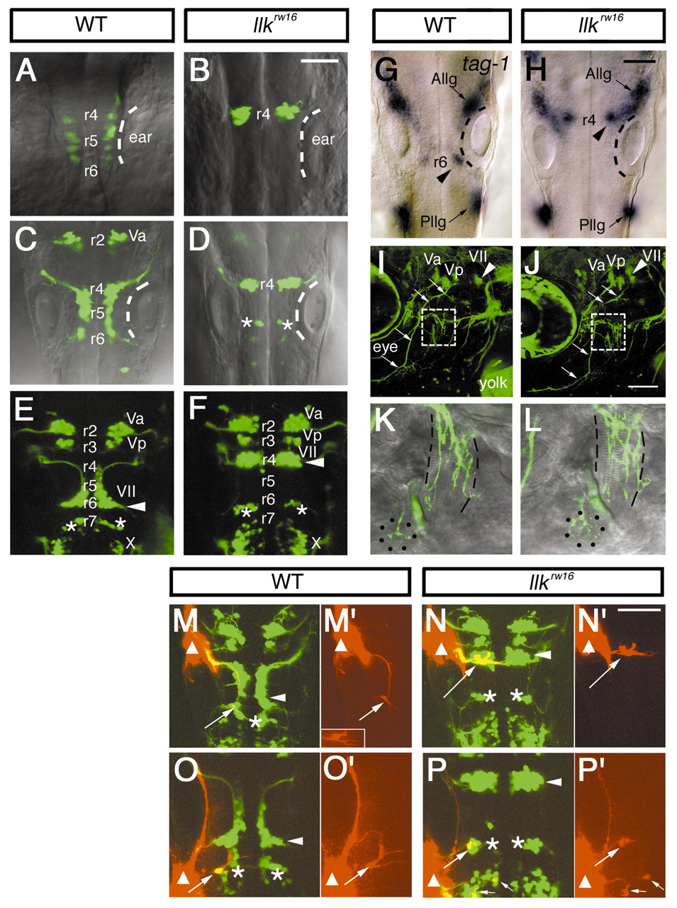
Fig. 3 Migration of the nVII motor neurons is specifically impaired in llk embryos. A-F, Isl1-GFP expression in the wild-type (A,C,E) and llk rw16 homozygous embryos (B,D,F) at 18 hpf (A,B), 24 hpf (C,D), and 48 hpf (E,F). In the wild-type siblings, the nVII motor neurons arise in r4, migrate caudally through r5 into r6 and form the nucleus in r6 (E, arrowhead). In contrast, in the llkrw16 embryos the GFP-expressing cells that arise in r4 fail to migrate and form an ectopic nucleus in r4 (F, arrowhead). Asterisks (D,E,F) indicate r6-derived putative OLe neurons, which migrate into r7 in the wild-type embryos. These neurons also fail to migrate in the mutant embryos and remain in r6 (D,F). (G,H) tag-1 mRNA expression in the wild-type (G) and llk rw16 (H) embryos at 24 hpf. tag-1-positive cells are located in r4 in the llkrw16 embryo (H, arrowhead). Dorsal views. The position of the ears are indicated by the broken lines. Va, Vp, anterior and posterior trigeminal nuclei, respectively; VII, facial nucleus; X, vagus nucleus; Allg, Pllg, anterior and posterior lateral line ganglion, respectively. (I-L) Isl1-GFP expression in the wild-type (I,K) and llk rw16 (J,L) embryos at 5 dpf. Arrowheads indicate the facial motor nucleus. The trajectories of facial motor axons are normal in the llk rw16 embryo (arrows). The axons reach the target organs (K,L, higher magnifications of the boxed regions in I and J). Lateral line organs and the cranial muscles are indicated by dots and broken lines, respectively. Lateral views; anterior is to the left. (M-N) The putative OLe neurons (arrows) projecting to the lateral lines are retrogradely labeled (red) in the wild-type (M,O) and llk rw16 (N,P) embryos. DiI was applied to the anterior (M,N) or posterior lateral line ganglion (O,P). Sites of DiI application are indicated by triangles. Small arrows indicate the vagal (X) motor neurons, which were labeled with DiI that diffused from the application site (P). Single-channel images of the labeled neurons are shown in M′, N′, O′ and P′. These neurons extend dendrites to the contralateral side of the brain. Inset in M′ shows another example in which dendritic processes were clearly labeled. Asterisks indicate r6-derived r7-located neurons, which fail to migrate and remain in r6 in the llk embryos. Arrowheads indicate the facial motor nuclei. Dorsal views. Scale bar: 50 µm.