
|
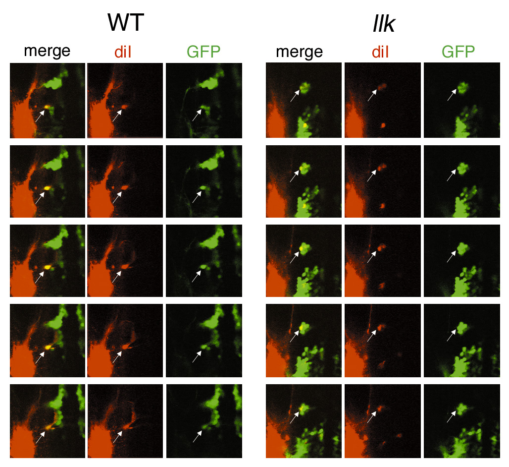
Fig. S1 The putative OLe neurons projecting to the posterior lateral lines are retrogradely labeled in the wild-type and llk rw16 embryos. Original serial optical sections, from which stack images were synthesized for Fig. 3O,P. Images were obtained by confocal microscopy at 10 mm interval. Diameters of confocal pinholes to detect the red and green signals were identical (154 mm) to avoid mis-identification. In each focal plane, co-localization of DiI and GFP signals are shown (arrows). Thus, we conclude that some of the GFP-expressing neurons extend to the posterior lateral lines, and we referred to these cells as ?putative OLe neurons? in the text.