
|
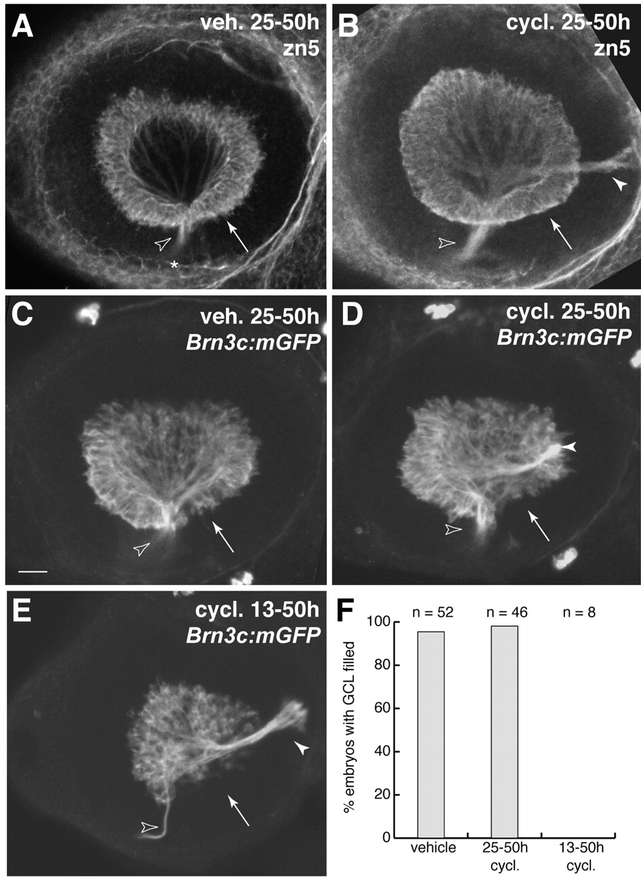
Fig. 9 Hedgehog signaling during retinal neurogenesis is not necessary for RGC differentiation. (A-D) Blockade of Hh signaling with cyclopamine at the start of retinal neurogenesis – 25 hpf – does not affect RGC formation. Two different RGC-specific markers, the zn5 antigen (A-B) and a Brn3c:mGFP transgene (C-D) were assessed at 50 hpf. In both vehicle-treated and cyclopamine (25-50 hpf)-treated animals, RGCs filled the entire GCL, including temporal retina (arrows), by 50 hpf. Note that, in wild type (A,C), all RGC axons left the eye via the optic disc (open arrowheads), whereas in cyclopamine animals (B,D) some RGC axons formed an ectopic fascicle projecting to the posterior of the eye (closed arrowheads). (E) Treatment with cyclopamine from 13-50 hpf delays RGC genesis. Note absence of GFP-expressing RGCs from the ventral half of the temporal retina (arrow). Zn5 staining gave similar results (not shown). Axon pathfinding defects were again observed (closed arrowhead). (F) Quantification of the phenotypes shown in A-E. Data were pooled over several experiments. Nearly all the vehicle and 25-50 hpf cyclopamine animals were finished with RGC genesis by 50 hpf, as shown by even filling of the GCL with RGCs. In the 13 hpf cyclopamine group, by contrast, none of the animals had a full GCL. Anterior/nasal is left and dorsal is up in all panels. Open arrowheads mark the optic disc/optic nerve head in all panels. A-E are z-projections of confocal stacks taken through the depth of the GCL. Scale bar: 25 µm. cycl., cyclopamine; veh., vehicle.