
|
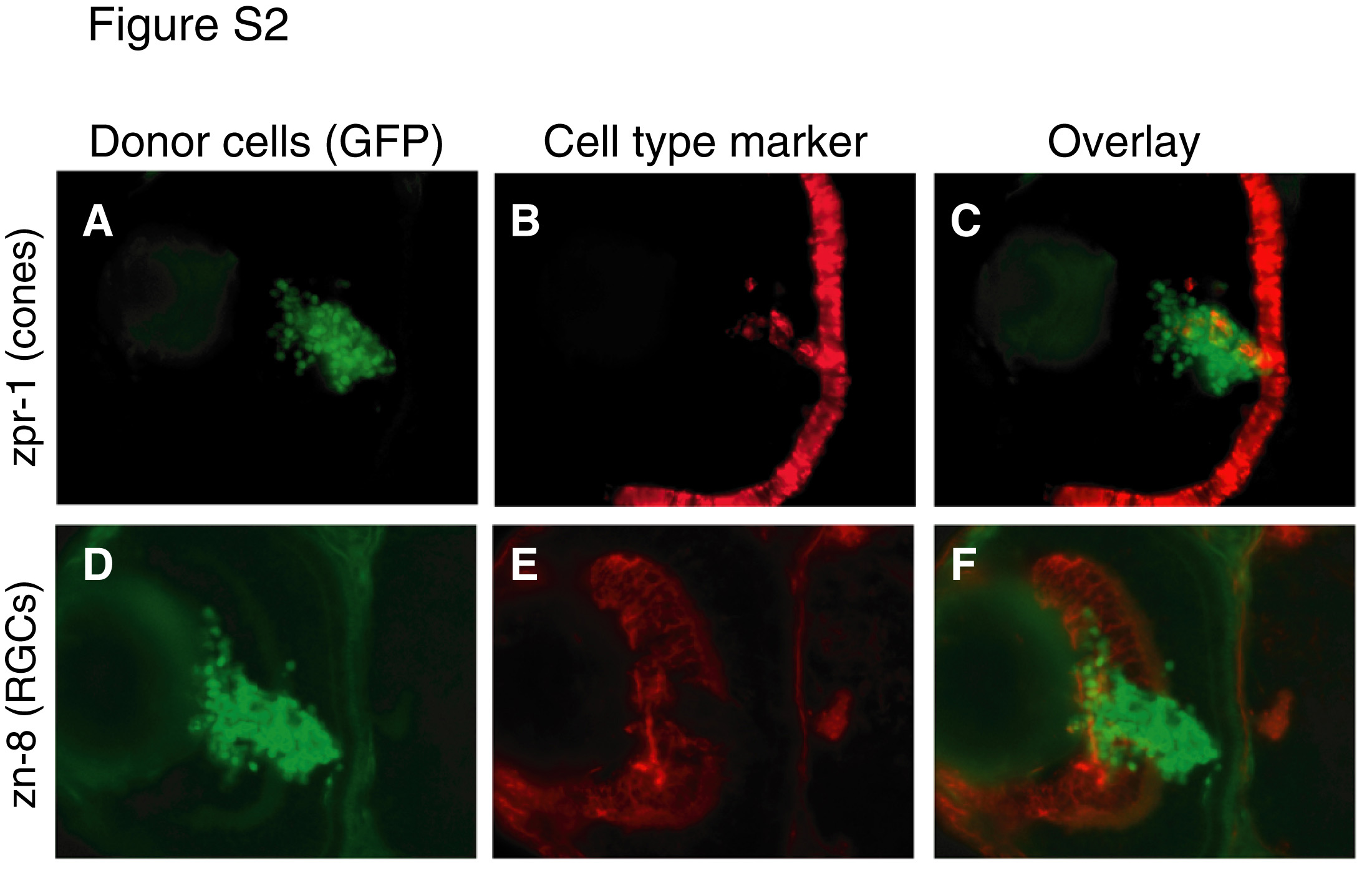
Fig. S2 Retinoblasts grafted into host retina differentiate and express RGC and photoreceptor markers. Retinal cells from 30 hpf histone 2A:GFP transgenic fish donors were transplanted into 30 hpf non-transgenic host retina. At 72 hpf, hosts were sectioned and immunostained using zpr-1, a marker of red/green cone photoreceptors (A-C), or zn-8, a marker of RGCs (D-F). Cell type marker analysis was performed on adjacent sections. (A,D) GFP donor cells, green. (B,E) Cell type markers, red. (C,F) Overlay showing expression of cell type markers by donor cells. All hosts examined (n=10) contained zn-8+ and zpr-1+ donor-derived cells. However, many donor cells failed to express either marker (C,F). The percentage of grafted cells in the GCL that failed to differentiate was estimated to be 65-85%.