
|
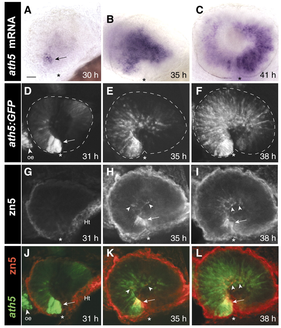
Fig. 1 The spatiotemporal pattern of RGC neurogenesis in zebrafish. Spread of ath5 mRNA (A-C), ath5:GFP (D-F) and RGC differentiation (G-I) across the retina. (A-C) Whole-mount embryos stained with an antisense ath5 riboprobe. (D-I) Whole-mount embryos double-stained with anti-GFP (D-F) and zn5 (G-I) antibodies. (J-L) Merge of D-F (ath5:GFP, green) with G-I (zn5, red), showing that the ath5 wave leads the RGC differentiation wave by several hours. D-L are z-projections of stacks of confocal images. Arrows in A,D and H-L indicate the ventronasal patch. No RGCs are present at 31 h (G), although zn5-immunoreactive tissue is seen in the heart (Ht). Arrowheads in H-I and K-L indicate zn5+ RGCs. Note that ath5 RNA expression is transient, becoming downregulated behind the wave (C), whereas GFP expression (F) persists longer and thus acts as an indelible marker of all cells that have expressed ath5. Asterisks mark the location of choroid fissure, which delineates the boundary between nasal and temporal retina. Anterior (nasal) is left and dorsal up in all figures. Scale bar: 25 µm. oe, olfactory epithelium; Ht, heart.