
|
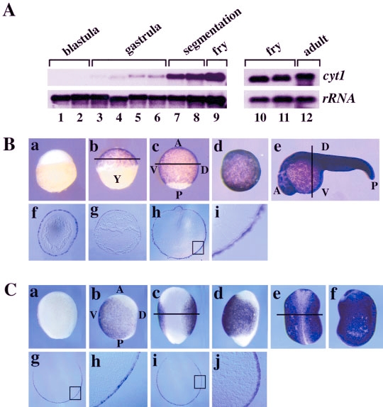
Fig. 2 The zebrafish cyt1 gene and the Xenopus XK81 gene are expressed in outer epithelial layers. A: Northern blot analysis of cyt1 expression. A cyt1 probe made from clone 1:4 (see Experimental Procedures section) was hybridized to Northern blots containing 1 μg of total RNA from various stages of development: Lane 1, 1-cell stage (3 hours postfertilization [hpf]); lane 2, sphere (4 hpf); lane 3, 50% epiboly (5 hpf); lane 4, 75% epiboly (8 hpf); lane 5, 90% epiboly (9 hpf); lane 6, tail bud (10 hpf); lane 7, 18 somites (18 hpf); lane 8, prim5 (24 hpf); lanes 9 and 10, long pec (48 hpf); lane 11, fry (1 week); lane 12, adult (3 month). Hybridization to a probe for 18S rRNA was used as a loading control. B: Whole-mount in situ hybridization analysis of cyt1 expression. A cyt1 probe (see Experimental Procedures section) was hybridized to zebrafish embryos from various stages: a, late blastula, sphere stage, 4 hpf; b, early gastrula, shield stage, 6 hpf; c, late gastrula, 85% epiboly, 9 hpf; d, mid-somitogenesis, 5-somite stage, 12 hpf; e, end of somitogenesis, prim 5, 24 hpf. Some embryos were sectioned after in situ hybridization to further define the expression of cyt1. f, section through embryo in b; g, section through embryo in c; h, section through embryo in e; i, enlargement of boxed region in h. The level of each section is indicated by a black line. Anterior is to the top in a–d and to the left in e. Dorsal is to the right in b–d and to the top in e. Dorsal cannot be assigned at the stage in a (sphere stage, 4 hpf). Dorsal is to the top in f–i. Y, yolk. C: Whole-mount in situ hybridization of XK81 expression. An XK81 probe (see Experimental Procedures section) was hybridized to Xenopus embryos from various stages; a and b, late gastrula, st. 12.5; c and d, mid-neurula, st. 15; e and f, tail bud, st. 20. Embryos were sectioned to further define XK81 expression: g, section through an early neurula stage embryo (st.14) at the level indicated in c; h, enlargement of area boxed in g; i, section through a late neurula stage embryo (st.18) at the level indicated in e; j, enlargement of boxed area in i. Anterior is to the top in a–f. a, c, and e are dorsal views. b, d, and f are lateral views. Dorsal is to the top in g–j. D, dorsal; V, ventral; A, anterior; P, posterior.