
|
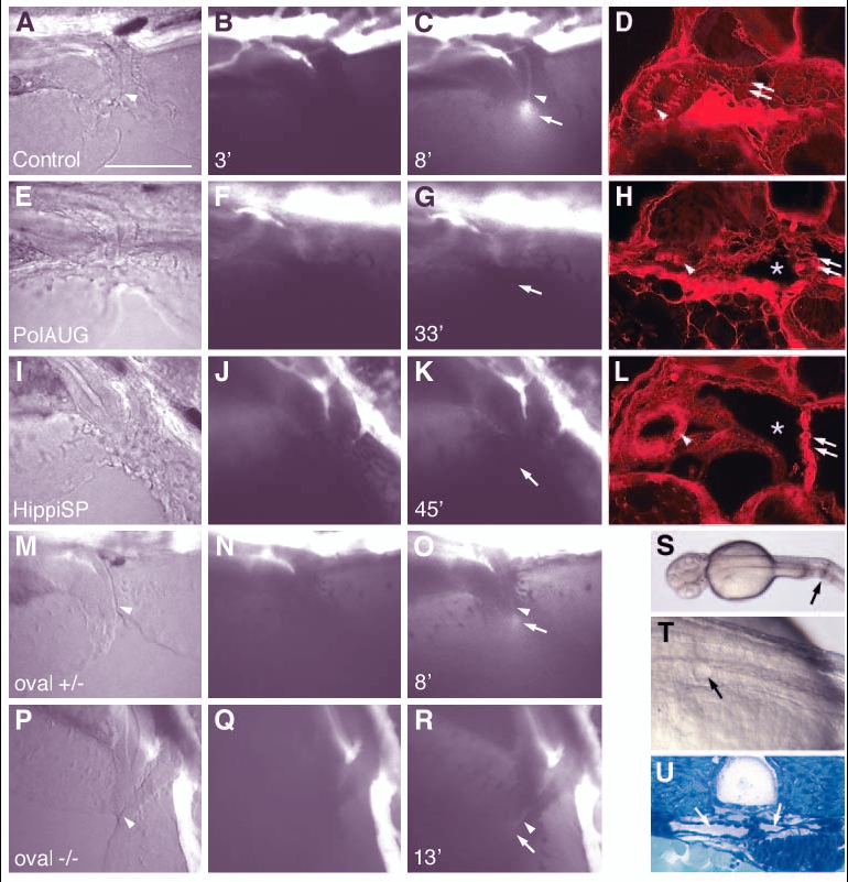
Fig. 4 Fluid flow is impaired by lack of normal cilia movement in the pronephros. Living embryos were injected with 5% tetramethylrhodamine-conjugated 70 k MW dextran into the circulation. After passage of the pronephric kidney, the dye was excreted at the cloaca (C, arrow). The images of the first column (A,E,I,M,P) are transmitted light images. The images in the second column (B,F,J,N,Q) were taken at 2-3 minutes post-injection, while the images in the third column (C,G,K,O,R) were captured when maximum excretion was reached. The time after injection that the image was captured is indicated in the bottom left of each panel. No fluorescent dye excretion via the cloaca was observed at timepoints >30 minutes in polarisAUG morphants (n=9) or hippiSP morphants (n=9), whereas in the control embryos excretion was observed in 22 individuals (n=27). On histology of the same embryos, all showed endocytic uptake of the dye in anterior duct cells (arrowhead) (D,H,L), indicating that the dye had been filtered via the glomerulus (double arrows) into the cyst lumens (*). In oval heterozygous embryos, excretion started at 5.3±0.4 seconds (n=2) (O), and 3 out of 5 oval homozygous embryos showed weak dye excretion at 13.7±5.5 seconds (n=3), whereas 2 did not show a visible output. In A-R anterior is to the left and dorsal is to the top. Mechanical obstruction of the pronephric ducts close to the cloaca (S, arrow) causes cystic distension of the anterior pronephric tubules within 30 minutes (T, arrow). The dilated tubules/glomerulus can be seen in cross sections (U, arrows). Scale bar: 100 µm.