
Image
|
Figure Caption
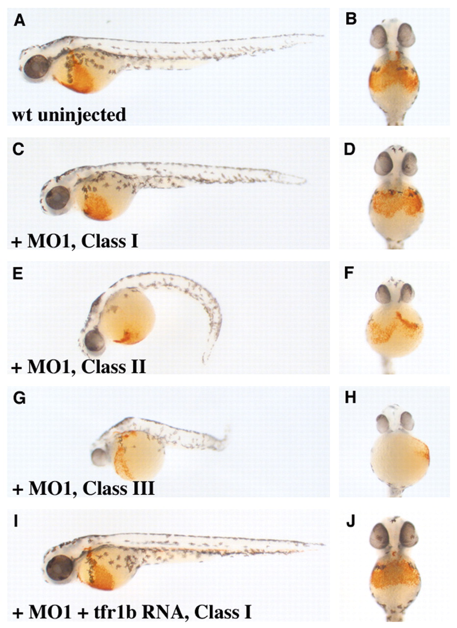
Fig. 7 Functional analysis of zebrafish tfr1b using morpholinos. (A,C,E,G,I) All show lateral views of 48 hpf o-dianisidine stained embryos, anterior to the left, with (B,D,F,H,J) showing ventral views of the same embryos. (A) Uninjected wild type. (C-H) Wild-type embryos injected with tfr1b MO1 exhibit three categories of phenotypic classes: (C,D) Class I embryo; (E,F) Class II embryo; and (G,H) Class III embryo. (I,J) Embryo co-injected with tfr1b MO1 and tfr1b cRNA.
Figure Data
Acknowledgments
This image is the copyrighted work of the attributed author or publisher, and
ZFIN has permission only to display this image to its users.
Additional permissions should be obtained from the applicable author or publisher of the image.
Full text @ Development