
|
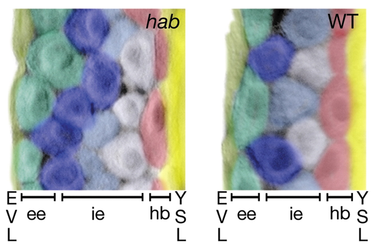
Fig. 5 Cell shape asymmetry and organization of the epiblast layer in hab mutants and wild-type embryos at 90% epiboly, showing Nomarski DIC photomicrographs of transverse sections of the lateral equator of fixed embryos stained with anti-ß-catenin. Cells are pseudo-colored to distinguish cells and layers are labeled at the bottom of the figure. At this stage, the mesoderm is only one cell layer thick, and endoderm cells have moved out of the plane of the section (Warga, 1999). ee, exterior layer of the epiblast layer; hb, hypoblast layer; ie, interior layer of the epiblast layer; YSL, yolk syncytial layer.