
|
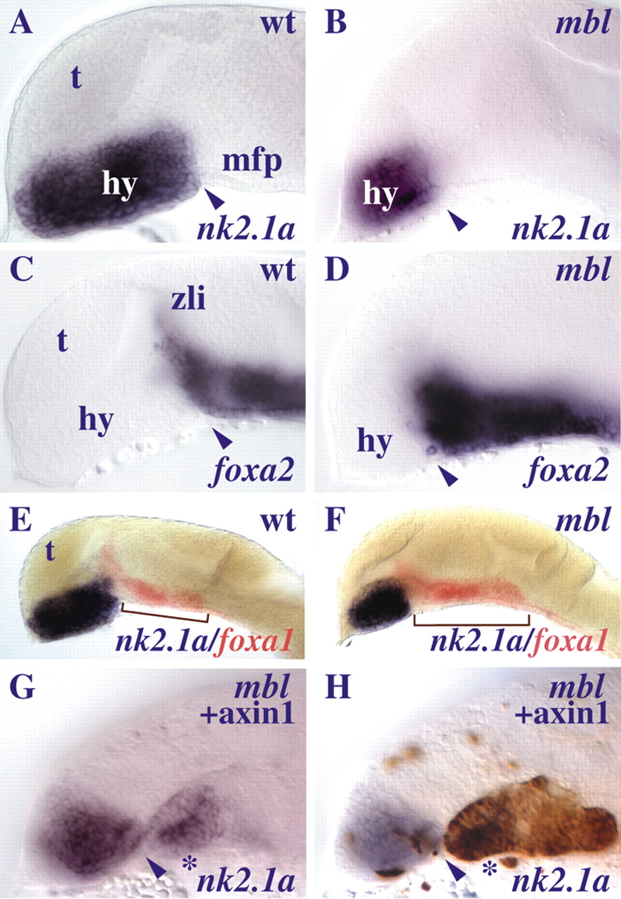
Fig. 2 mbl embryos show a reduction in the size of the hypothalamus coupled with an expansion in posterior diencephalic/midbrain floorplate. Lateral views of brains of embryos between 25 and 31 somites with anterior to the left. (A-F) The mbl embryos (B,D,F) show reduced hypothalamic tissue and rostral expansion of floorplate-expressed genes compared to the wild-type embryos (A,C,E). Arrowheads point at the rostral limit of floorplate marker expression (A-D). Brackets show the AP expansion of diencephalic/midbrain foxa1 expression (red) in the mbl (F) compared to the wild-type embryo (E). (G-H) Transplanted cells overexpressing axin1 (brown) restore nk2.1a expression (blue, asterisk) in the rostral ventral neural tube of an mbl embryo. Abbreviations: hy, hypothalamus; mfp, medial floorplate; t, telencephalon; zli, zona limitans intrathalamica; wt, wild type.