
|
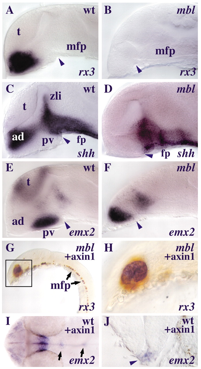
Fig. 5 mbl embryos lose rostral but retain caudal hypothalamic marker gene expression. (A-H) Lateral views of brains of embryos at about 31 somites with anterior to the left. (A-D) The hypothalami of the mbl embryos (B,D) do not express rx3 (B) and lose the anterior domain of shh expression (D) compared to the wild-type embryos (A,C). (E-F) The mbl embryo (F) retains expression of the hypothalamic marker emx2 which is expressed in the caudal hypothalamus of the wild-type embryo (E). Arrowheads point at the caudal limit of the hypothalamus. (G-H) Transplanted cells overexpressing axin1 (brown) that have incorporated into the rostral ventral CNS restore rx3 expression (blue) in the mbl embryo (G). (H) A higher magnification view of the transplant (boxed) of this embryo. (I-J) Some Axin1-overexpressing cells that incorporate into the floorplate domain of a wild-type host ectopically express emx2 (arrows). The transverse section (indicated by the left arrow in I) shows ectopic emx2 expression in the floorplate (arrowhead in J). Abbreviations: ad, anterior-dorsal hypothalamus; fp, floorplate; mfp, medial floorplate; pv, posterior-ventral hypothalamus; t, telencephalon; zli, zona limitans intrathalamica.