
|
OPTICS: MAGNIFICATION: DATE OF IMAGE: SUBMITTER COMMENTS:
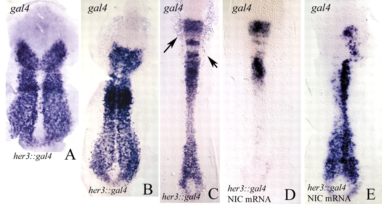
Fig. 2 (A-C) Flat preparations of her3::gal4 embryos (tail-bud, A; two-somite stage, B; six-somite stage, C) labelled by in situ hybridisation with a gal4 probe. The gal4 pattern closely corresponds to the endogenous her3 transcription pattern. Compare with Fig. 1. The arrows in C indicate areas of gal4 expression in epidermal tissues. In cranial regions of the developing spinal cord, reporter mRNA persists for longer than do the transcripts of the endogenous her3 gene (see Fig. 1). (D,E) Two examples of her3::gal4 embryos injected with mRNA for notch1a-intra and probed for gal4. Note that transcription of her3::gal4 is repressed. Anterior is towards the top.
| Preparation | Image Form | View | Direction |
| not specified | still | not specified | not specified |