
|
OPTICS: MAGNIFICATION: DATE OF IMAGE: SUBMITTER COMMENTS:
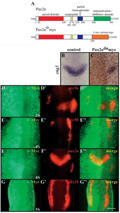
Fig. 4 Pax2a and Pax8 are required for induction of otic cell fates. (A) Schematic map of protein domain structure of Pax2a and the dominant-negative variant, dnPax2a-myc. In dnPax2a-myc, the 295 N-terminal amino acids are fused in frame with six Myc-epitopes. The numbers indicate amino acids of the domains (Lun and Brand, 1998). (B,C) dnPax2a-myc acts in a dominant-negative fashion. At the three-somite stage, eng3 (blue) is expressed in the isthmic region (B), whereas in wild-type embryos injected with dnpax2a-myc, the side expressing the transgene shows reduced or no eng3 (blue) expression (C). The distribution of the Myc-epitope (brown) indicates the localization of the Pax2a variant in nuclei. Expression of dominant-negative Pax2a leads to downregulation of sox9a (D,D′,D′′), sox9b (E,E′,E′′) and pax2a (F,F′,F′′) but has only slight effects on dlx3b (G-G′). (D-G) Fluorescence images of a-Myc antibody labeling; (D′-G′) fluorescence images of mRNA in situ hybridization probes; (D′′-G′′) merged fluorescence images. (B-G′′) Dorsal views, anterior towards the top. Scale bar: 120 μm.
| Preparation | Image Form | View | Direction |
| not specified | still | not specified | not specified |