
Image
|
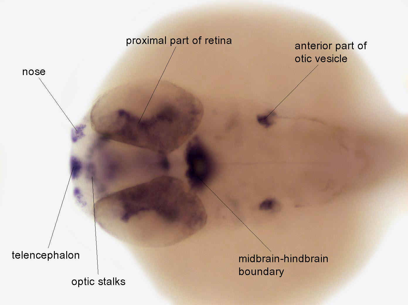
Figure Caption
Fig. 6 At 36 hrs, expression is observed in the ganglion cell layer of the retina, the adenohypophysis, and the hyoid arch. Expression has disappeared from the posterior otic vesicle and the somites. An increase of expression is observed in median fin fold
Developmental Stage
Prim-15 to Prim-25
Orientation
| Preparation | Image Form | View | Direction |
| whole-mount | still | dorsal | anterior to left |
Figure Data