|
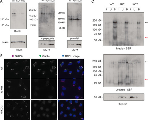
Procollagen processing in giantin KO MC3T3 cells. (A) Immunoblots of cell lysates taken from WT and giantin KO MC3T3 cells. Antibodies used are as indicated. (B) Single-plane widefield images of WT and giantin KO MC3T3 cells immunolabeled for giantin (green) and the cis-Golgi marker GM130 (magenta). The nucleus is stained in DAPI. Scale bar = 10 µm. (C) Immunoblots of a GFP trap of WT and giantin KO MC3T3 cell cultures transiently expressing GFP-COL1A1. Media and lysates were assayed after 24 h of ascorbate treatment, which was conducted 24 h after transfection. The input (I), unbound (U), and bound (B) fraction of each pull-down is shown probed with either SBP or tubulin antibodies as indicated underneath each blot. Black arrows indicate full-length procollagen, and red arrows the N-propeptide.
|