
Fig. 7
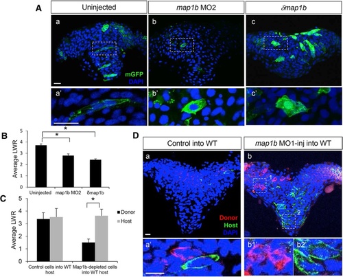
Map1b functions cell autonomously in the neural ectoderm. a Hindbrain sections of 4–5 som uninjected (a, a′), map1b MO2-injected (b, b′) and δmap1b-injected (c, c′) embryos mosaically-expressing mGFP (green). Nuclei are labeled in blue with DAPI. (a′–c′) Higher magnification of boxed areas in (a–c) respectively. Scale bars: 20 µm. b Quantification of the LWR of cells in 4–5 som uninjected, map1b MO2-injected and δmap1b-injected embryos. (*) Indicates statistical significance (P <0.01 for uninjected vs map1b MO2 and P <0.001 for uninjected vs δmap1b-injected) using a Kruskal-Wallis test followed by Dunn’s post-hoc test. c Quantification of the LWR of control donor cells vs host WT cells and map1b MO1-injected donor cells vs host WT cells. (*) Indicates statistical significance (P <0.0001) using Student’s T-test. d Hindbrain sections of 4–5 som WT hosts mosaically-expressing mGFP and transplanted with (a) mRFP-labeled control donor cells or (b) mRFP-labeled map1b-MO1 donor cells. Nuclei are labeled with DAPI (blue). (a′, b1′ and b2′) Higher magnifications of boxed areas in (a, b1 and b2) respectively. Scale bars: 10 µm |
| Fish: | |
|---|---|
| Knockdown Reagent: | |
| Observed In: | |
| Stage: | 1-4 somites |