
Fig. 6
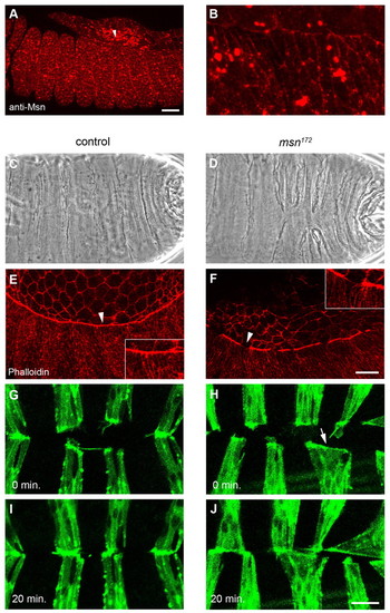
Drosophila misshapen is required for actin-based cell constriction and segment alignment during dorsal closure. (A,B) Anti-Msn antibody staining of a stage 13 control embryo, lateral view with anterior to the left. Arrowhead in (A) indicates the region shown in a magnified view in (B). Large spots probably represent non-specific signal. (C,D) Cuticle preparations of control (C) and msn172 mutant embryos (D) after dorsal closure completion. (E,F) Phalloidin (F-actin) staining of stage 14 control (E) and msn172 mutant embryos (F). Arrowheads demarcate regions shown as insets. (G-J) Time-lapse analysis of the final stages of dorsal closure in control (G,I) and msn172 mutant embryos (H,J) expressing GFP-Actin in the engrailed domain. Embryos are shown at time points 0 min (G,H) and +20 min (I,J). Scale bars: in A, 25 μm for A,B; in F, 25 μm for E,F; in J, 10 μm for G-J. |