
- Title
-
The Developmental Brain Gene NPAS3 Contains the Largest Number of Accelerated Regulatory Sequences in the Human Genome
- Authors
- Kamm, G.B., Pisciottano, F., Kliger, R., and Franchini, L.F.
- Source
- Full text @ Mol. Biol. Evol.
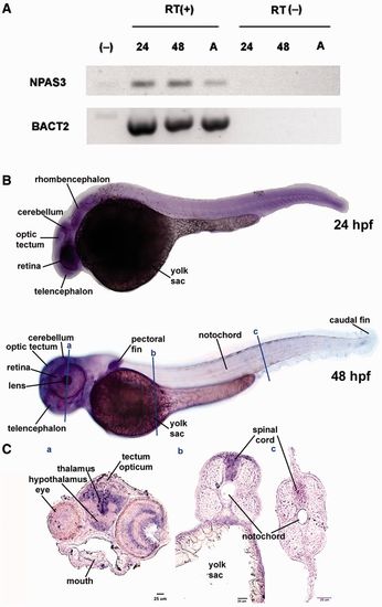
Expression of the NPAS3 gene during zebrafish development. (A) RT-PCR analysis of NPAS3 expression in zebrafish embryos and adult brain. RT-PCR products of NPAS3 and control gene b-actin2 are shown: (-), negative control; 24, 24 hpf; 48, 48 hpf; A, adult brain with (+) or without (-) reverse transcriptase. (B) Whole mount in situ hybridization studies show the spatial expression pattern of the gene NPAS3 at 24 hpf (top) and 48 hpf (bottom). (C) In situ hybridization on coronal cryostat slices showing a detail of the NPAS3 expression pattern in 48 hpf zebrafish embryos. |
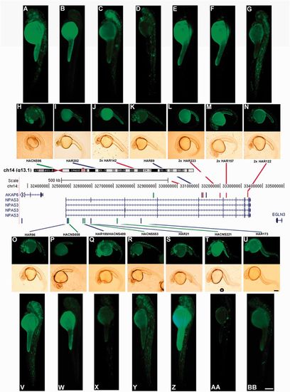
The NPAS3 gene contains the largest number of HAEs. In the central panel, schematic drawing modified from the UCSC genome browser, showing the NPAS3 locus in genomic context on chromosome 14q13.1. Below, the four splicing variants of the NPAS3 gene are shown. The coloured bars indicate the location of the 14 HAEs. Blue bars indicate HARs (Pollard, Salama, King, et al. 2006; Pollard, Salama, Lambert, et al. 2006); green bars indicate HACNS (Prabhakar et al. 2006); and red bars show 29-mammals HARs (2× HAR; Lindblad-Toh et al. 2011). The overlapping HAE is shown by a green–blue bar. On top and below of the central panel, fluorescent and bright field microphotographs showing representative transgenic zebrafish of each HAE expressing EGFP at 24 hpf (H, I, J, K, L, M, N, O, P, Q, R, S, T, and U) and 48 hpf (A, B, C, D, E, F, G, V, W, X, Y, Z, AA, and BB). Small boxes (U and BB) represent 250 μm. |
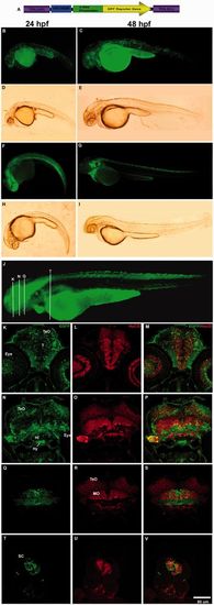
HACNS96 drives EGFP expression to the developing nervous system in stable and transient transgenic zebrafish lines at 24, 48, and 72 hpf. (A) Diagram of the transgene structure based on the Tol2 system. Fluorescent (B, C, F, and G) and bright field (D, E, H, and I) photomicrographs of selected whole mount transient or stable transgenic zebrafish embryos expressing the reporter protein EGFP under control of the human HACNS96 sequence at 24 hpf (B, D, F, and H) and 48 hpf (C, E, G, and I). (J) Representative transgenic zebrafish expressing EGFP under the control of HANCS96 at 72 hpf where white bars indicate approximately the location of coronal cryostat sections showing a detail of the EGFP expression pattern (K, N, Q, and T), cells expressing the early neuronal marker HuC/D (L, O, R, and U) and the resulting overlay (M, P, S, and V). TeO, tectum opticum; T, midbrain tegmentum; DT, dorsal tegmentum; Po, preoptic region; MO, medulla oblongata; OC, otic capsule; SC, spinal cord; Hi, hypothalamus; Hy, hypophysis. EXPRESSION / LABELING:
|
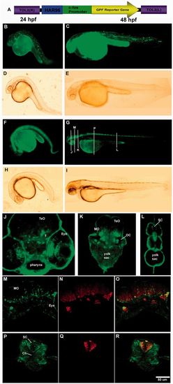
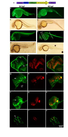
EGPF expression driven by HAR96 in stable and transient transgenic zebrafish lines at 24 and 48 hpf. (A) Diagram of the transgene structure based on the Tol2 system. Lower panel, fluorescent (B, C, F, and G) and bright field (D, E, H, and I) photomicrographs of selected whole mount transient or stable transgenic zebrafish embryos expressing the reporter protein EGFP under control of the human HAR96 sequence at 24 hpf (B, D, F, and H) and 48 hpf (C, E, G, and I). Coronal cryostat sections showing a detail of the EGFP expression pattern at three levels (white vertical bars in G) of the 48 hpf developing zebrafish (J, K, and L). Below, cells expressing EGFP (M and P) the early neuronal marker HuC/D (N and Q) and the resulting overlay (O and R) in the 48 hpf developing zebrafish (sections levels are approximately indicated in G). TeO, tectum opticum; T, midbrain tegmentum; DT, dorsal tegmentum; Po, preoptic region; MO, medulla oblongata; OC, otic capsule; SC, spinal cord; Hi, hypothalamus; Hy, hypophysis. |

Differences in expression driven by human and chimpanzee HAR202 orthologs. (A) Diagram of the transgene structure based on the Tol2 system. Below, fluorescent (B, C, F, and G) and bright field (D, E, H, and I) photomicrographs of selected whole mount transgenic zebrafish embryos expressing the reporter protein EGFP under control of the human (HAR202-Hs) or the chimpanzee (HAR202-Pt) sequences at 24 and 48 hpf. Coronal cryostat sections showing a detail of cells expressing EGFP (J, M, P, and S), cells expressing the early neuronal marker HuC/D (K, N, Q, and T) and the resulting overlay (L, O, R, and U) in the 48 hpf developing zebrafish (sections levels are approximately indicated in G). TeO, tectum opticum; T, midbrain tegmentum; DT, dorsal tegmentum; Po, preoptic region; MO, medulla oblongata; OC, otic capsule; SC, spinal cord; Hi, hypothalamus; Hy, hypophysis. |
|

Unillustrated author statements |