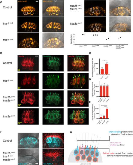
Tall and short hair cells of the lateral crista display biases for Tmc proteins in mechanotransduction. (A) Representative confocal images of 4-Di-2-ASP (orange) dye uptake assays showing differential dependencies of tall and short hair cells on Tmc proteins in the lateral cristae of 7-dpf larvae. Solid lines outline tall hair cells with 4-Di-2-ASP uptake in a wild-type sibling, a tmc2acwr3 single mutant, and a tmc2bcwr2tmc2acwr3 double mutant. In each wild-type tall hair cell, there is an accumulation of dye near the cell’s base. The dashed lines outline tall hair cells with diminished 4-Di-2-ASP uptake in the tmc1cwr5 single mutant and the tmc1cwr4 single mutant or short hair cells with no or little 4-Di-2-ASP uptake in the tmc2bcwr2tmc2acwr3 double mutant and the tmc2acwr3 single mutant. Graph of lengths of hair cells that have diminished 4-Di-2-ASP uptake. Each data point represents a single cell’s length and associated genotype. Cells associated with each data point are shown in confocal images with dashed outlines. (B) Representative confocal images of FM 4–64 (red) uptake assays for a heterozygous sibling control (tmc2bcwr2/+tmc2acwr3/+, B’), a tmc1cwr4 single mutant (B”), a tmc2bcwr2tmc2acwr3 double mutant (B”’), and a tmc2acwr3 single mutant (B””), each at 7-dpf. The lateral crista hair cells express GCaMP6s-CAAX (green), making each cell apparent. The red or white asterisks mark the short hair cells with or without FM4-64 loading, respectively. Red, yellow, or white arrowheads mark tall hair cells with normal, reduced, or no apparent FM4-64 dye loading, respectively. Red asterisk in panel (B”’) marks a short hair cell erratic that permits FM4-64 entry in the absence of Tmc2a and Tmc2b. (C–E) Graphs of normalized hair cell fluorescence intensity ratios (ITall HCs (hair cells)/IShort HCs, C) and the percentages of hair cells that loaded with FM4-64 (nloaded HCs/ntotal HCs, D,E). (C) Two-tailed unpaired student’s t-test, ****P < 0.0001. (D) One-way ANOVA, P = 0.6076. (E) Two-tailed unpaired student’s t-test, ****P < 0.0001, **P = 0.0017. For the tmc1cwr4 single mutant, nTall HCs = 23, ncrista = 4. For the tmc2bcwr2tmc2acwr3 double mutant, nTall HCs = 15, nshort HCs = 28, ncrista = 3. For the tmc2acwr3 single mutant, nTall HCs = 21, nshort HCs = 30, ncrista = 3. For the controls, nTall HCs = 27, nshort HCs = 40, ncrista = 4. (F) 4-Di-2-ASP (left, live) and FM1-43FX (right, fixed with hair bundles labeled by phalloidin) uptake assays demonstrate that mechanotransduction of lateral crista hair cells is absent in the tmc2bcwr8tmc1cwr4tmc2acwr6 triple mutant, regardless of hair cell subtype. Control, tmc2bcwr8/+tmc1cwr4/+tmc2acwr6/+. (A,B,F) Scale bar = 5 μm. (G) Model of Tmc dependencies in the central thickness of the lateral crista.
|
