
Fig. 5
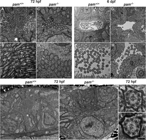
pam−/− zebrafish exhibit microvillar and ciliary assembly defects in the pronephros. Transmission electron micrographs of transverse sections through the pronephros of pam−/− embryos and their wildtype siblings at 72 hpf (A–D and I,J) and 6 dpf (E–H); the images shown were taken from approximately midway along the pronephros. At 72 hpf, the pronephros lumen of wildtype zebrafish is occluded with numerous cilia surrounded by a dense array of microvilli (A,B). Although densely packed cilia were evident in the pronephric lumen of pam−/− embryos at this stage, brush border microvilli were absent (C,D). At 6 dpf, the lumen of the wildtype zebrafish pronephros was more open; numerous cilia appeared in the lumen, which was surrounded by epithelial cells extending a dense array of microvilli (E,F). In contrast, the lumen of the pam−/− pronephros showed a severe reduction in the number of cilia along with the continued absence of a brush border (G,H). Occasional cytosolic axonemes (inset in H) and undocked centrioles/basal bodies (arrow in G and see Supplemental Fig. S6) were observed in the epithelial cells lining the pronephric duct of the pam−/− embryos. Lower magnification images (I,J) of the region surrounding the pronephros of the 72 hpf embryos shown in panels (A,B) illustrate the general cellular architecture. The cilia present in pam−/− embryos at 72 hpf were morphologically normal (K,L). Bars = 100 nm (K,L), 500 nm (A,C,F,H), 250 nm (B,D), 1 μm (G) and 2 μm (E,I,J). |
| Fish: | |
|---|---|
| Observed In: | |
| Stage Range: | Protruding-mouth to Day 6 |