
Fig. 3
- ID
- ZDB-FIG-130826-17
- Publication
- Hollenbach et al., 2013 - Different Regulation of Physiological and Tumor Angiogenesis in Zebrafish by Protein Kinase D1 (PKD1)
- Other Figures
- All Figure Page
- Back to All Figure Page
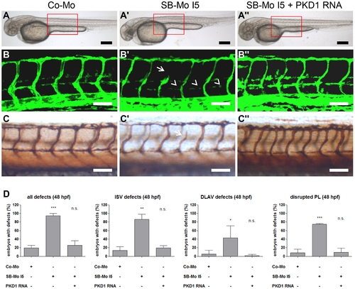
Injection of human PKD1 mRNA rescued vascular defects in PKD1 morphant tg(fli1:EGFP) zebrafish embryos. A–A3, Overall morphology of Co-Mo - injected (2 ng), PKD1 SB-Mo I5 - injected (500 pg) or of SB-Mo I5 and PKD1 mRNA (100 pg) co-injected fish embryos showed no morphological alterations. Red boxes indicate regions of pictures shown in (B–C3). B–C3, Embryos were analyzed at 48 hpf by confocal microscopy (B–B3) or anti-GFP antibody staining (C–C3). Morpholino-based expression silencing of PKD1 in zebrafish disrupted partially formation of the vasculature (ISVs (arrows), PL (arrowheads)) (B2, C2). Coinjection of SB-Mo I5 (500 pg) and PKD1 mRNA (100 pg) rescued the vascular malformation in PKD1 morphants (B3, C3). D, Quantification of the number of embryos with vascular defects, ISV defects, DLAV defects and disrupted PL at 48 hpf. For each group at least 40 embryos were analyzed. Black scale bars: 300 μm; white scale bars: 100 μm. *P<0.05, **P<0.01, ***P<0.001. |