
- Title
-
Specification of hematopoietic and vascular development by the bHLH transcription factor SCL without direct DNA binding
- Authors
- Porcher, C., Liao, E.C., Fujiwara, Y., Zon, L.I., and Orkin, S.H.
- Source
- Full text @ Development
|
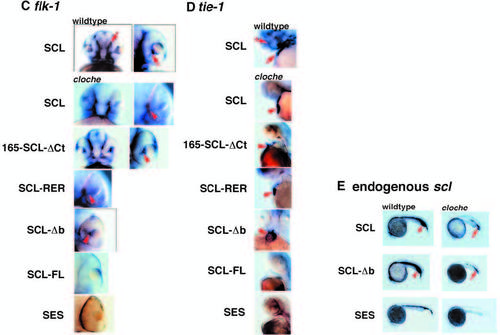
DNA-binding domain by SCL is not required for initiation of primitive erythropoiesis and vasculogenesis in cloche. Embryos are shown in lateral views and oriented with rostral (left) and caudal (right) in A,B and E; in ventral view with rostral (top) in C; and in lateral views with rostral (top) in D. Embryos are shown at 48 hpf in A-D, and at 24 hpf in E. Wild-type embryos microinjected with full-length murine SCL are shown on the top of A-D. cloche embryos microinjected with murine SCL, 165- SCL-ΔCt, SCL-RER, SCL-Δb, SCL-FL and SES are shown in that descending order in A-D. (A) o-dianisidine staining of SCL-injected wild-type embryo revealed abundant blood cells pooling in the cardinal sinus (arrowhead) and major vessels of the trunk (arrowhead with *). SCL, 165-SCL- ΔCt, SCL-RER and SCL-Δb rescued blood in cloche, where the blood cells pooled in the trunk (arrowhead) and tail (arrowhead with *). SCL-FL and SES failed to rescue blood cells in cloche, where the trunk was devoid of blood cells and the tail contained only 5-10 cells (arrowhead with *). The 5-10 cells observed in SCL-FL and SES injected embryos were also seen in uninjected controls (data not shown). (B) Same results of blood rescue as observed in A, except blood cells were detected by alas-e riboprobe. Rescued blood cells are indicated (arrowheads). (C) flk-1 in situ detects cranial vasculature in wild-type embryos (arrowheads with *). Rescue of choroid plexus vessels abutting the optic disc were consistently observed with injections of SCL, 165-SCL-ΔCt, SCL-RER and SCL-Δb (arrowheads). SCL-FL and SES failed to rescue endothelial cells in cloche and the embryos lacked flk-1 staining. (D) tie-1 in situ delineates cranial vasculature (arrowhead with +) and endocardium (arrowhead with *) of wild-type embryos. Microinjections with SCL, 165- SCL-ΔCt, SCL-RER and SCL-Δb rescued endocardial cells and tie-1 expression in cloche mutants (arrowheads), but did not rescue tie-1 in the cranial vasculature. SCL-FL and SES failed to rescue tie-1 in cloche. (E) Endogenous SCL expression was induced by ectopic expression of mouse SCL and SCL-Δb. The domain of endogenous SCL expression was expanded in wild-type embryos (arrowheads) and rescued in cloche mutants (arrowheads with *). SES microinjection did not induce endogenous SCL expression. 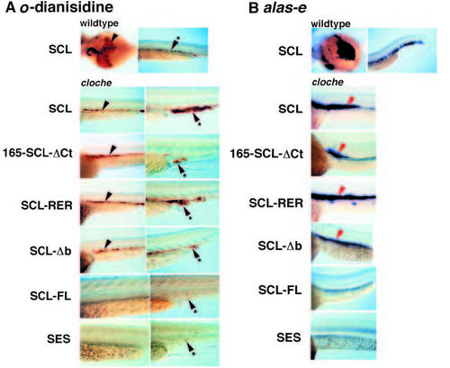
|