
- Title
-
Somatotopy of the lateral line projection in larval zebrafish
- Authors
- Alexandre, D. and Ghysen, A.
- Source
- Full text @ Proc. Natl. Acad. Sci. USA
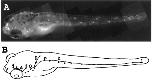
(A) Vitally labeled neuromast hair cells in a 6-day-old zebrafish larvae. The dye also is taken up by the nasal epithelium. (B) Neuromasts and lateral line nerve branches whose projection was examined in the present work. Neuromasts on one side of the fish are drawn as small circles. The nerve branches that were not back-filled in this work are not shown. A, anterior lines; P, midbody posterior line; D, dorsal line; O, occipital line; 1, last two neuromasts of the posterior midbody line; 2, second neuromast of the posterior midbody line; 3, neuromast located dorso anterior to the ear vesicle; 4, last neuromast of the supra orbital line. Neuromasts 1 and 2 are innervated by the posterior lateral line ganglion, and neuromasts 3 and 4 are innervated by the anterior lateral line ganglion. Magnification: x20. |
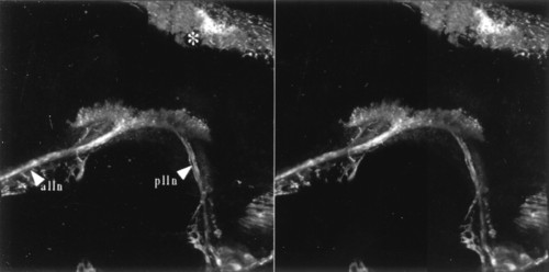
Vitally labeled lateral line primary projection. Stereo pair of a dorso lateral view seen from the outside of the animal. Anterior is to the left and dorsal to the top. alln, anterior lateral line nerve; plln, posterior lateral line nerve. * indicates dorsal head skin. Magnification: x160. |
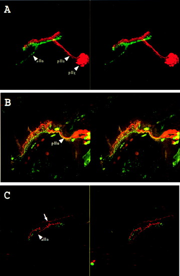
Double labeling of the lateral line system. (A) Labeling of the midbody posterior lateral line nerve (plln) with rhodamine-dextran (red) and of the anterior lateral line nerve (alln) with fluoresceine-dextran (green). Stereo pair of a side view seen from the outside of the animal. Anterior is to the left and dorsal to the top. pllg, posterior lateral line ganglion. The anterior lateral line ganglion could not be imaged because of the limited working distance of the lens. (B) Labeling of the afferents of the second neuromast of the midbody posterior lateral line (2 in Fig. 1) with fluoresceine-dextran and the last two neuromasts of the same line (1 in Fig. 1) with rhodamine-dextran. Stereo pair of a dorsolateral view seen from the outside of the animal. Anterior is to the left and dorsal to the top. plln, posterior lateral line nerve. (C) Labeling of the afferents of a neuromast located dorso anteriorly to the ear (3 in Fig. 1) with rhodamine-dextran and the last neuromast of the supra orbital line (4 in Fig. 1) with fluoresceine-dextran. Stereo pair of a dorsolateral view seen from the outside of the animal. Anterior is to the left and dorsal to the top. alln, anterior lateral line nerve. The arrow indicates trigeminal descending fibers, which often were labeled in these preparations. Magnification: x160. |
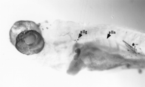
Labeling of the afferents of the second neuromast of the midbody posterior lateral line (numbered 2 in Fig. 1 and Table 1). * indicates site of dye application. Magnification: x60. |