
- Title
-
The cloche and spadetail genes differentially affect hematopoiesis and vasculogenesis
- Authors
- Thompson, M.A., Ransom, D.G., Pratt, S.J., MacLennan, H., Kieran, M.W., Detrich,III, H.W., Vail, B., Huber, T.L., Paw, B., Brownlie, A.J., Oates, A.C., Fritz, A., Gates, MA., Amores, A., Bahary, N., Talbot, W.S., Her, H., Beier, D.R., Postlethwait, J.H., and Zon, L.I.
- Source
- Full text @ Dev. Biol.
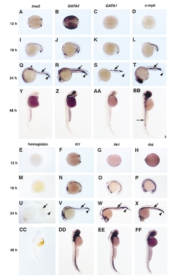
Comparison of the expression patterns of hemoglobin and seven different genes involved in hematopoiesis and/or vasculogenesis in developing zebrafish embryos. Whole-mount in situ hybridization staining of wild-type zebrafish embryos with lmo2 (A, I, Q, Y); gata2 (B, J, R, Z); gata1 (C, K, S, AA); c-myb (D, L, T, BB); fli1 (F, N, V, DD); flk1 (G, O, W, EE); flt4 (H, P, X, FF). Hemoglobin revealed by o-dianisidine staining of embryos (E, M, U, CC). Ages of embryos are 12 h (A–H), 18 h (I–P), 24 h (Q–X), and 48 h (Y–FF). Embryos shown in A–H are viewed dorsally with anterior to the left. At 12 h lmo2 (A), gata2 (B), and fli1 (F) are expressed in cells located in two strips of ventral mesoderm. Embryos in I–FF are viewed laterally with either the anterior to the left (I–X) or at the top (Y–FF). By 18 h, the ventral mesodermal cells are converging to form the ICM. All seven genes are expressed in converging cells at this time. Just prior to circulation, around 24 h, ICM cells expressing the seven genes and hemoglobin (U) are located at the midline between the gut and the notochord (arrow in Q–X). In addition cells in the tail express lmo2 (Q), gata2 (R), fli1 (V), flk1 (W), and flt4 (X), but not gata1 (S), c-myb (T), or hemoglobin (U) (arrowhead, Q–X). At this time, lateral cells in the trunk region around the pronephric primordia and dorsal aorta express lmo2 (asterisk in Q). In 48-h-old embryos, the embryonic red blood cells are in circulation and putative definitive blood cell progenitors that express c-myb (arrow in BB) are developing in the dorsal aorta. At 48 h lmo2 (Y), gata2 (Z), and flt4 (FF) are not expressed in hematopoietic cells, while gata1 (AA) expression is decreasing, and fli1 (DD), flk1 (EE), and hemoglobin (CC) continue to be highly expressed. All embryos, except in U are in 70% glycerol/PBS. U is in BB/BA. EXPRESSION / LABELING:
|
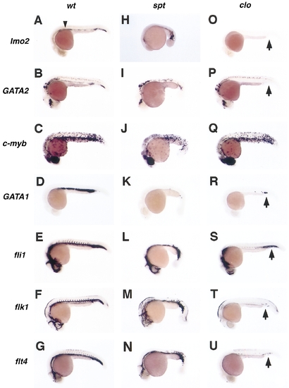
Comparison of the expression patterns of seven genes involved in hematopoiesis and vasculogenesis in wild-type embryos and embryos homozygous for one of two different mutations. Whole-mount in situ hybridization of 24-h-old wild-type (A–G), spadetail (sptb104) (H–N), and cloche (clom39) (O–U) embryos with lmo2 (A, H, O), gata2 (B, I, P), c-myb (C, J, Q), gata1 (D, K, R), fli1 (E, L, S), flk1 (F, M, T), and flt4 (G, N). Arrowhead in A indicates trunk lmo2 expression. In spt mutant embryos, cells expressing lmo2 (H) and gata2 (I) are found in the morphologically distorted ICM region and tails. However, very few if any hematopoietic cells expressing gata1 (K) or c-myb (J) are seen in these embryos. The expression levels of the vascular markers fli1 (L), flk1 (M), and flt4 (N) appear normal in these embryos; however, the patterns are distorted by the abnormal somites. In clo mutant embryos, ICM cells and tail cells that express lmo2 (O) or gata2 (P) are not found. While the expression of fli1 (S), gata1 (R), c-myb (Q), flk1 (T), and flt4 (U) is missing in the anterior ICM of clo mutant embryos, a small number of posterior ICM and tail cells continue to express these markers (arrows in O–U). The expression of c-myb in the eye, gut, and skin-associated cells appears normal in all of these mutants (C, J, Q). EXPRESSION / LABELING:
|
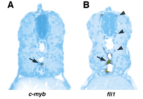
The relative positions of cells that express c-myb or fli1 in 48-h-old embryos. Cross sections through the trunk of 48-h-old embryos stained for c-myb (A) expression or fli1 (B) expression by whole-mount in situ hybridization. The positive cell in A (arrow) lies in the ventral wall of the dorsal aorta. In B, staining is seen in the endothelial cells (arrow) of the dorsal aorta and caudal vein as well as in regions coinciding with the segmental artery and vein (arrowheads). EXPRESSION / LABELING:
|
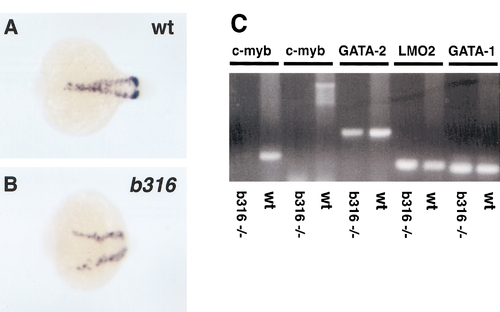
Deletion of the c-myb gene in b316 mutants does not result in a loss of gata1 expression in zebrafish embryonic erythroid cells. Whole-mount in situ hybridization of wild-type (A) and b316 (B) embryos at approximately 18 h postfertilization with gata1. Note that the b316 embryo development is arrested at about 5-somites, but that it appears to have a normal level of gata1 expression. PCR of DNA from wild-type or b316 embryos with two different c-myb primer pairs, and with primers specific for gata2, lmo2, and gata1 (C). c-myb is not amplified with either primer pair from DNA made from b316 embryos, while it is amplified from DNA made from wild-type embryos. gata2, lmo2, and gata1 all amplify in both b316 and wild-type embryos. EXPRESSION / LABELING:
|
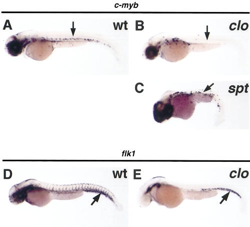
The cloche (clom39) and spadetail (sptb104) mutations affect definitive progenitors in the dorsal aorta. Whole-mount in situ hybridization of 48-h-old wild-type (A, D), clom39 (B, E), and sptb104 (C) embryos with c-myb (A–C) and flk1 (D, E). All three mutations greatly reduce the number of cells that express c-myb in the dorsal aortas of 48-h-old embryos (arrows A–C). Cells expressing flk1 are seen in the tails of 48-h-old embryos (arrows D, E). EXPRESSION / LABELING:
|
Reprinted from Developmental Biology, 197, Thompson, M.A., Ransom, D.G., Pratt, S.J., MacLennan, H., Kieran, M.W., Detrich,III, H.W., Vail, B., Huber, T.L., Paw, B., Brownlie, A.J., Oates, A.C., Fritz, A., Gates, MA., Amores, A., Bahary, N., Talbot, W.S., Her, H., Beier, D.R., Postlethwait, J.H., and Zon, L.I., The cloche and spadetail genes differentially affect hematopoiesis and vasculogenesis, 248-269, Copyright (1998) with permission from Elsevier. Full text @ Dev. Biol.