
- Title
-
Neuronal and neuroendocrine expression of lim3, a LIM class homeobox gene, is altered in mutant zebrafish with axial signaling defects
- Authors
- Glasgow, E., Karavanov, A.A., and Dawid, I.B.
- Source
- Full text @ Dev. Biol.
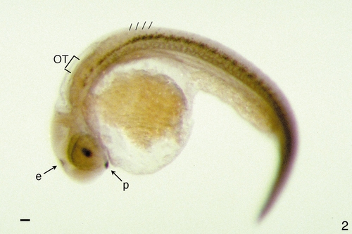
Expression of Lim3 is limited to the pituitary, epiphysis, hindbrain, and ventral spinal cord in 28-h zebrafish embryos. The expression of Lim3 protein in zebrafish was determined by whole-mount immunohistochemistry using anti-Lim3 antibody. At 28 h postfertilization, Lim3 protein expression is restricted to nuclei of the pituitary anlage (p), the epiphysis (e), the hindbrain, and the spinal cord. OT, otic vesicle. The approximate posterior borders of the first four somites are indicated with hash marks. The staining in the eye lens is artifactual, due to nonspecific trapping of the antibody. Scale bar is 100 μm. EXPRESSION / LABELING:
|
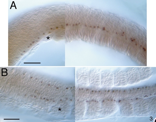
Expression of Lim3 protein in 10-somite-stage zebrafish embryos. (A and B) Ten-somite-stage embryos stained as whole mounts were dissected from the yolk and flatted under a coverslip. Anterior is to the left. Three to four Lim3-positive cells are seen in each segment associated with each somite. The first somite is indicated by an asterisk. Scale bars are 50 μm. (A) Lateral view, dorsal is up. Lim3-positive cells are in a ventral position in the hindbrain and spinal cord. (B) Dorsal view. The Lim3-positive cells in the hindbrain appear to be continuous with those of the spinal cord. EXPRESSION / LABELING:
|
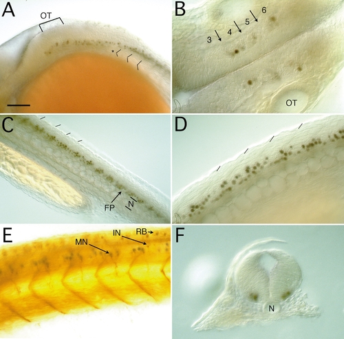
Expression of Lim3 in the hindbrain and spinal cord in 28-h embryos. Anterior is to the left except in F. (A) Lateral view of the hindbrain and anterior spinal cord. Dorsal is up. Lim3-positive cells in the hindbrain extend to rhombomere 4, just anterior to the otic vesicle, OT (brackets). Two rows of Lim3-positive cells are seen in the posterior hindbrain, one row appearing to be continuous with the ventral spinal cord cells. The first somite is marked with an asterisk, and the first three somite borders are indicated. (B) Ventral view of the hindbrain. Three clusters of Lim3-positive cells are seen in the center of rhombomeres 4, 5, and 6. Rhombomeres 3–6 are numbered, with arrows indicating the location of the rhombomere borders. OT, otic vesicle. (C) Lateral view of the spinal cord. Lim3-positive cells are arranged segmentally along the ventral spinal cord. The plane of focus is at a slight angle so that superficial structures are anterior and deeper structures are posterior. At the anteriormost side, somite boundaries are clearly seen (hash marks). Next, the Lim3-positive cells come into focus slightly above the midline in the ventral spinal cord. At the midline, the floor plate (FP) and notochord (N) are in focus. Toward the posterior, the Lim3-positive cells on the deeper side of the midline come into focus. (D) Closeup lateral view of the spinal cord. The stained nuclei of the segmentally arranged cells are clearly seen. (E) Lateral view of the spinal cord, double labeled with anti-Lim3 (purple) and zn12 (brown) antibodies. The zn12 monoclonal antibody recognizes the L2/HNK1 epitope on many neurons. Cells with Lim3-stained nuclei also have ventrally projecting axons that stain with zn12, whereas not all cells with zn12-labeled ventrally projecting axons express Lim3. An example of a double-labeled ventrally projecting motoneuron (MN) is indicated, along with a probable interneuron (IN) that is negative for Lim3 and positive for zn12. The soma and axons of the dorsally located Rohon–Beard (RB) sensory neurons label heavily with zn12. The zn12 immunoreactive myosepta are prominent. (F) Cross section through the anterior spinal cord. The Lim3-positive cells are in a ventrolateral position on both sides of the midline well separated from the floor plate cells and the notochord (N). Scale bar is 100 μm in A and C, 50 μm in B, D, and E, and 60 μm in F. EXPRESSION / LABELING:
|
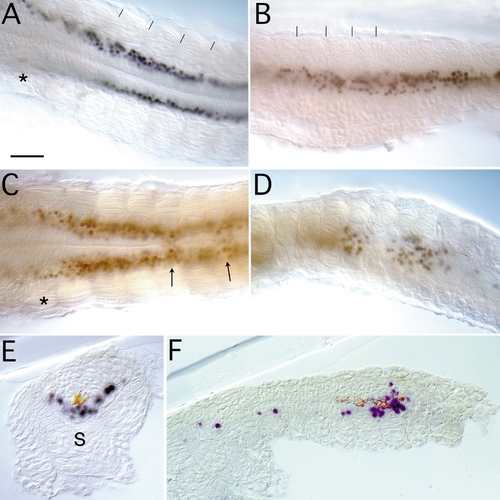
Expression of Lim3 in the spinal cord of no tail (ntl) and floating head (flh) mutant zebrafish. Twenty-eight-hour embryos were dissected from the yolk and flatted under a coverslip (A–D), or sectioned (E,F). Anterior is to the left, except in E. (A) Ventral view of the anterior spinal cord in a ntl mutant. There is clear separation of Lim3-positive cells across the midline. The first somite is marked with an asterisk and somite borders are indicated by hash marks. (B) Lateral view of ntl embryo at the level of the trunk. The Lim3-positive cells are restricted to the ventral spinal cord, but with a slightly broader distribution than in the wild type and an apparent loss of segmental organization. (C) Ventral view of an flh-mutant anterior spinal cord. The first somite is marked by an asterisk. An excess number and loss of organization of Lim3-positive cells is observed in the anterior spinal cord of the mutant embryos. Variably, around the level of the fifth somite, the midline separation of Lim3-positive cells breaks down (arrows). (D) Lateral view of flh embryo at the posterior end of the trunk. At this level, areas with unorganized clumps of Lim3-positive cells alternate with areas lacking Lim3-positive; in the tail of flh mutants there is no Lim3 expression. (E) Cross section of flh-mutant anterior spinal cord, double labeled with anti-Lim3 antibody (purple) and monoclonal antibody MZ15 which stains notochord and floor plate (brown). MZ15 staining is seen only on the ventricular surface of the spinal cord, and Lim3-positive cells are present across the midline. In flh mutants, axial mesodermal cells are transfated from notochord to somitic mesoderm (S). (F) Parasagital section of the posterior trunk region of a Lim3 (purple) and MZ15 (brown) double-labeled flh mutant embryo. Lim3 expression correlates with expression of MZ15. Scale bar is 50 μm in A–D and F, and 70 μm in E. EXPRESSION / LABELING:
|
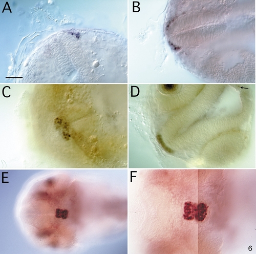
Expression of Lim3 in the pituitary anlage. Embryos were stained in whole mount, dissected from the yolk, and photographed from a ventral view. The embryos are oriented so that anterior faces left, except in A where anterior faces up. The plane of focus cuts through part of the ventral diencephalon. A–D, wild-type embryos; E and F, flh mutant embryos. (A) At the 21-somite stage, Lim3 is present in approximately 8 cells, laterally on the left side of the anteriormost end of the neural tube. (B) At the 24-somite stage, cells on both sides of the neural tube express Lim3 still in a lateral position. (C) By 28 h, Lim3-expressing cells have moved to the midline and coalesced into the pituitary cluster. (D) An anterior view of a 28-h embryo. The optical section runs from the anterior edge of the pituitary cluster through the epiphysis, with the diencephalon in the plane of focus. A stained cell can be seen slightly below the focal plane in the epiphysis (arrow). The Lim3-positive cells of the pituitary cluster are one cell thick at the anterior edge. The staining in the eye lens is not nuclear and is probably artifactual. (E,F) The same flh embryo shown at two magnifications; since the enlarged pituitary was not in one plane of focus at the higher magnification, F was assembled from two images of the same embryo (see E). When comparing the wild-type embryo in C with the mutant in F, it is apparent that the flh pituitary is twice as large in the A/P dimension, but slightly more compact in the L/R dimension; there are approximately twice as many Lim3-positive cells in the mutant than in the wild-type pituitary. Scale bar is 50 μm except in E where it is 100 μm. EXPRESSION / LABELING:
PHENOTYPE:
|
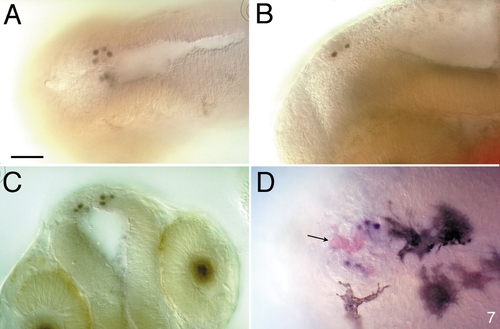
Expression of Lim3 in the epiphysis is restricted to a subset of projection neurons. (A–C) Four to five Lim3-expressing cells are seen on each side of the midline in a ventrolateral position in the epiphysis of a 28-h embryo. (A) Dorsal view. (B) Lateral view. (C) Anterior view. This optical section through the epiphysis, with the diencephalon in focus, is slightly deeper than in Fig. 5D. (D) Dorsal view of a 2-day-old embryo. Whole-mount embryos were double labeled with anti-S-antigen (red) and anti-Lim3 (blue, nuclei). Four to five projection neurons express Lim3 protein. Several cells, especially in the center of the epiphysis, express the photoreceptor marker, S-antigen (arrow). By this stage several prominent melanocytes are seen. Scale bar is 50 μm. |

Unillustrated author statements EXPRESSION / LABELING:
|
Reprinted from Developmental Biology, 192, Glasgow, E., Karavanov, A.A., and Dawid, I.B., Neuronal and neuroendocrine expression of lim3, a LIM class homeobox gene, is altered in mutant zebrafish with axial signaling defects, 405-419, Copyright (1997) with permission from Elsevier. Full text @ Dev. Biol.