
- Title
-
Mutations affecting development of zebrafish digestive organs
- Authors
- Pack, M., Solnica-Krezel, L., Malicki, J., Neuhauss, S.C., Schier, A-, F., Stemple, D.L., Driever, W., and Fishman, M.C.
- Source
- Full text @ Development
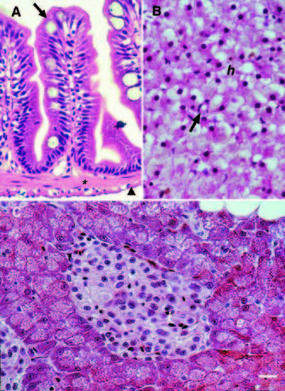
Histology of adult zebrafish digestive organs. (A) Cross section of intestine. Arrow points to intestinal epithelium, arrowhead to outer longitudinal smooth muscle; * indicates inner circular smooth muscle. (B) Liver. Arrow points to intrahepatic bile duct. (C) Pancreas. e, exocrine cells; h, hepatocytes; i, islet. Scale bars, A, 25 μm; B and C, 15 μm. |
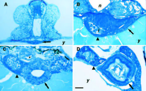
Histology of the digestive organs in the developing zebrafish. Cross sections of proximal gut. (A) A small lumen is visible in the developing intestine (arrow) of the 36 hour postfertilization (hpf) embryo. (B) The intestine has enlarged (arrow) and the pancreatic islet (i) and exocrine precursor cells (arrowhead) are visible in the 52 hpf embryo. (C) The intestinal epithelium (arrow) has begun to polarize in the 72 hpf larvae. Exocrine precursors surround the islet (arrowhead). (D) The intestinal epithelium (arrow) has fully polarized and the pancreatic exocrine cells have differentiated (arrowhead) at 4 dpf. The liver is rostral to the plane of section in A and B. e, exocrine pancreas; i, pancreatic islet; l, liver; n, notochord; y, yolk; * labels pneumatic duct of gas bladder. Scale bars, A and D, 25 μm; B, 20 μm; C, 30 μm. |
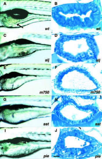
Mutations affecting the developing zebrafish intestine. (A,C,E,G,I) Lateral views of living 5 dpf larvae. (B,D,F,H,J) Corresponding histological cross-sections through the mid-intestine. Lumen size varies with position along the anterior-posterior axis and with muscular contractions. (A,B) Wild type. The intestinal epithelium (arrow) is highly folded, bile is visible in the lumen and the gas bladder is inflated. The intestinal epithelium is polarized with basal nuclei and a microvillus brush border. (C,D) slj m74, (E,F) m750, (G,H) sstm311, (I,J) piem497. The intestinal wall of all mutants is thin, lacks folds and the epithelium is not fully polarized and shows signs of degeneration. Scale bar, B, F and H, 20 μm; D, 15 μm; J, 25 μm. |
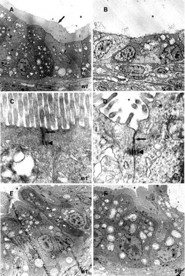
Electron micrographs showing regional intestinal differentiation. (A,C,E) wild type and (B,D,F) sljm74. (A) The wild-type anterior intestinal epithelium is columnar and has a prominent microvillus brush border (arrow). (B) sljm74 anterior intestinal epithelial cells are cuboidal with few microvilli. (C,D) Adherens (arrowhead) and tight junctions (arrow) are present in the anterior intestine of both (C) wild type and (D) sljm74. (E,F) Posterior intestinal epithelium appears normal in both (E) wild type and (F) sljm74. * labels the intestinal lumen. Magnification: A, x950; B, x1100; C, x22,000; D, x28,500; E, x2200; F, x2950. |
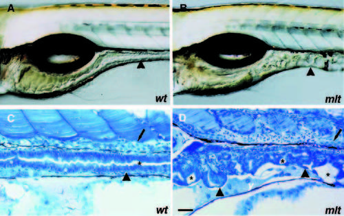
mltm498 perturbs posterior intestine. (A,C) Wild type, (B,D) mel. The posterior intestinal epithelium is disorganized and the mesenchyme expanded. * labels the intestinal lumen; arrowheads, posterior intestine; small arrows, pronephric duct. Scale bars, C and D, 30 μm. PHENOTYPE:
|
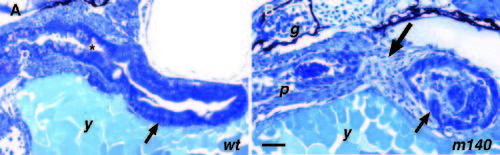
In m140 the esophagus fails to develop normally. (A) wild type, saggital section, 4 dpf. The intestine (arrow) is joined to the pharynx by the esophagus (*). (B) m140, 4 dpf. There is no connection (large arrow) between the intestine (small arrow) and pharynx, both of which have begun to degenerate. p, pharynx; g, glomerulus; y, yolk. Scale bars: A, 40 μm; B, 30 μm. PHENOTYPE:
|
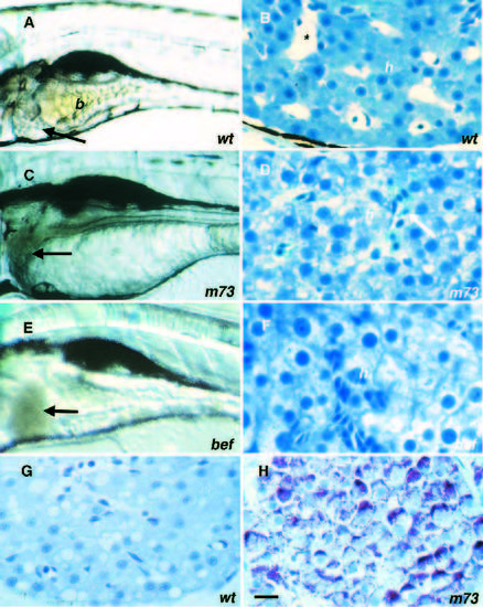
Liver mutants. (A,BG) Wild type. (C,D,H) m73. (E,F) bef m362. (A) Lateral view of wild-type 5 dpf larva with bile (b) in intestinal lumen; arrow points to liver. (B) Cross section of wild-type liver 5 dpf with erythrocytes in the sinusoids (*) and hepatocytes (h). (C) The day-4 m73 liver (arrow) has a gray-brown hue. (D) m73 hepatocytes are degenerating and erythrocytes (arrow) are pooling in the sinusoids. (E) befm362 liver (arrow) has a red-brown hue. (F) befm362 hepatocytes also degenerate and compress the sinusoids. (G) Wild-type liver 6 dpf does not stain with PAS. (H) m73 liver 6 dpf stains for PAS. Scale bar, 15 μm. |
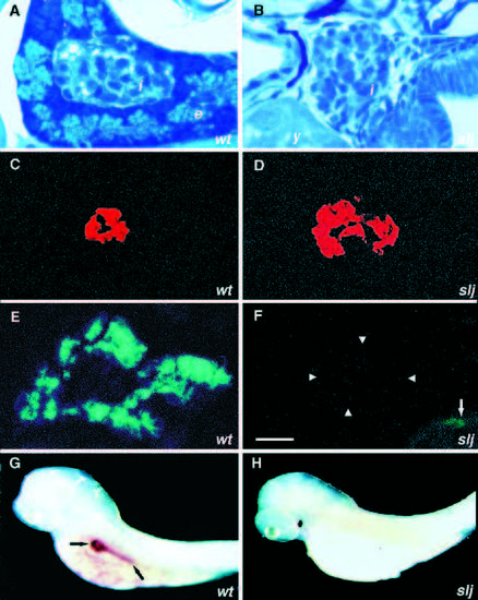
Pancreas mutants. Pancreas development in (A,C,E,G) wild type and (B,D,F,H) sljm74 embryos. (A) Cross-section of the pancreas at 4 dpf showing islet (i) and exocrine (e) cells. (B) In sljm74 the islet appears normal but the exocrine cells have not developed. (C) Immunoreactive insulin in the wild-type islet. (D) Immunoreactive insulin in the sljm74 pancreas. (E) Immunoreactive carboxypeptidase A in the wild-type exocrine pancreas. (F) There is little if any immunoreactive carboxypeptidase A in the slj pancreas at 4 dpf. Arrrowheads mark the position of islet cells immunoreactive for insulin. Arrow, background staining in intestine. (G) GATA-5 in situ hybridization in the wild type shows expression in the exocrine pancreas (arrows). (H) sljm74 does not express GATA-5 in the exocrine pancreas. Scale bars, A,B,D and F, 15 μm; C,E, 20 μm. |

ZFIN is incorporating published figure images and captions as part of an ongoing project. Figures from some publications have not yet been curated, or are not available for display because of copyright restrictions. EXPRESSION / LABELING:
|
|
ZFIN is incorporating published figure images and captions as part of an ongoing project. Figures from some publications have not yet been curated, or are not available for display because of copyright restrictions. |