
- Title
-
Mutations affecting the development of the embryonic zebrafish brain
- Authors
- Schier, A.F., Neuhauss, S.C., Harvey, M., Malicki, J., Solnica-Krezel, L., Stainier, D.Y., Zwartkruis, F., Abdelilah, S., Stemple, D.L., Rangini, Z., Yang, H., and Driever, W.
- Source
- Full text @ Development
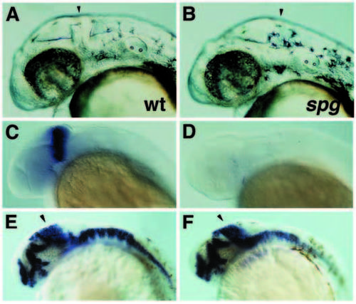
Phenotype of spiel ohne grenzen (spg) mutants on day 2 of development. (A,B) DIC image of wild-type (A) and spgm216 mutant (B) embryos at 30 hpf. Arrowhead indicates the position of midbrain-hindbrain boundary. (C,D) Expression of engrailed-2 in wild-type (C) and strong spgm216 mutant (D) embryos at 28 hpf. Weaker spgm216 mutants retain a small dorsal patch of engrailed-2 expression. (E,F) Expression of dlx2 and hlx1 in wild-type (E) and spgm216 mutant (F) embryos at 28 hpf. Arrowhead indicates position of the prospective tectum. |
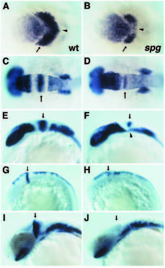
Phenotype of spiel ohne grenzen (spg) mutants during somitogenesis. (A,B) Expression of pax[zf-b] (arrow) in the region of the midbrain and presumptive midbrain-hindbrain boundary region and pax6 (anterior to pax[zf-b] stripe) in the forebrain of wildtype (A) and spgm216 mutant (B) embryos at the 1-somite stage; dorsal view. Note the reduced medial expression domain of pax[zf-b] (arrowhead). (C,D) Expression of pax[zf-b] at the midbrainhindbrain boundary (arrow) and pax6 (forebrain, eye anlage and hindbrain) in wild-type (C) and spgm216 mutant (D) embryos at the 10-somites stage; dorsal view. (E,F) Lateral view of embryos in C and D, respectively. Note the absence of ventral pax[zf-b] expression at the midbrain-hindbrain boundary and the shift of the pax6 expression domains in forebrain and hindbrain with respect to each other (arrowhead). (G,H) Expression of wnt1 in wild-type (G) and spgm216 mutant (H) embryos at the 14-somites stage. Note the reduction of wnt1 expression at the midbrain-hindbrain boundary (arrow). (I,J) Expression of pax[zf-b] (optic stalk, midbrainhindbrain boundary (arrow), hindbrain) in wild-type (G) and spgm216 mutant (H) embryos at 26.5 hpf. EXPRESSION / LABELING:
PHENOTYPE:
|
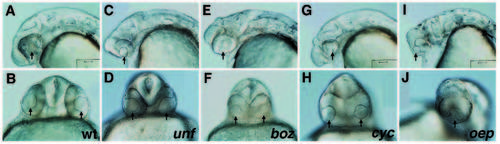
Phenotypes of mutations affecting the formation of ventral neuroectoderm on day 1 of development. Lateral (A,C,E,G,I) and anteriorventral (B,D,F,H,J) views of wild-type (A,B), uncle freddy (unf)m768 (C,D), bozozok (boz)m168 (E,F), cyclops (cyc)m122 (G,H) and one-eyed-pinhead (oep)m134 (I,J) mutant embryos at 28 hpf. Arrows indicate the position of the lens. PHENOTYPE:
|
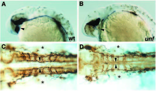
Phenotypic analysis of uncle freddy (unf)m768 mutants. (A,B) Expression of sonic hedgehog in hypothalamus (arrowhead) and floor plate in wild-type (A) and unfm768 mutant (B) embryos at 29 hpf. (C,D) Medial longitudinal fascicles (arrows), lateral longitudinal fascicles and commissures in hindbrain stained with antibody against acetylated tubulin in wild-type (C) and unfm768 mutant (D) embryos at 29 hpf. Note the partial fusion of the medial longitudinal fascicles at the midline of the unfm768 mutant embryo. Asterisks indicate the position of the otic vesicles. |
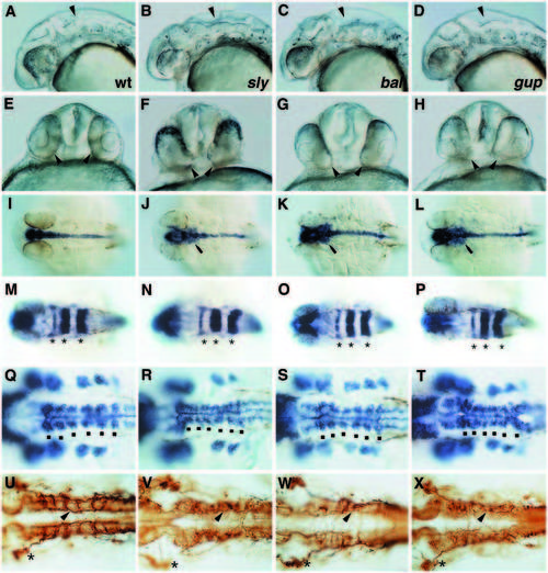
Phenotypes of mutations affecting notochord and brain. (A,E,I,M,Q,U) wild type; (B,F,J,N,R,V) sleepy (sly)m86; (C,G,K,O,S,W) bashful (bal)m190; (D,H,L,P,T,X) grumpy (gup)m189. Lateral (A,B,C,D) and anterior-ventral (E,F,G,H) view of embryos at 28 hpf. Note the enlarged hindbrain ventricles and the irregular morphology of the hindbrain (arrowhead in A-D) and the position of the eyes (arrowheads in EH). (I,J,K,L) Expression of sonic hedgehog at 29 hpf; dorsal view. Note the lateral expansion posterior to the eye in mutant embryos (arrow in J,K,L). (M,N,O,P) Expression of rtk1 in rhombomeres 1, 3 and 5 (asterisks). (Q,R,S,T) Expression orf dlx2 in pharyngeal arch primordia and hlx1 in hindbrain at 29 hpf; dorsal view. Note the slightly irregular expression domains of hlx1 adjacent to rhombomere boundaries (dots) in mutant embryos. (U,V,W,X) Medial longitudinal fascicles (arrowhead in the region of rhombomere 5), lateral longitudinal fascicles and commissures in hindbrain stained with antibody against acetylated tubulin at 29 hpf. Dorsal view. Asterisks highlight the position of the trigeminal ganglion. |
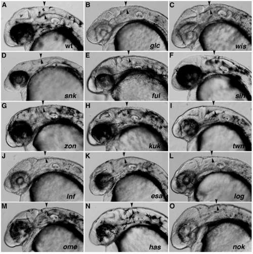
Phenotypes of mutations affecting ventricle enlargement. (A) Wild type; (B) glaca (glc)m309; (C) white snake (wis)m427; (D) snakehead (snk)m273; (E) fullbrain (ful)m133; (F) silent heart (sih)b109; (G) zonderzen (zon)m163; (H) kuehler kopf (kuk)m484; (I) turned down (twn)m359; (J) landfill (lnf)m551; (K) eraserhead (esa)m725; (L) logelei (log)m628/logm673 transheterozygous; (M) oko meduzy (ome)m98; (N) heart and soul (has)m129; (O) nagie oko (nok)m227 (Malicki et al., 1995a) between 30 and 33 hpf. Arrowheads outline the border of the hindbrain ventricle anterior to the otic vesicle (asterisk). PHENOTYPE:
|
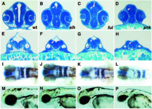
Phenotypic analysis of mutations affecting ventricle enlargement. (A,E,I,M) Wild type; (B,F,J,N) silent heart (sih)b109; (C,G,K,O) fullbrain (ful)m133; (D,H,L,P) snakehead (snk)m273. (A,B,C,D) Transverse sections through eye and lens at 28 hpf. (E,F,G,H) Transverse section through anterior ear and otolith at 28 hpf. Note the different degrees of ventricle (white arrowheads) reduction in sih, ful and snk. (I,J,K,L) Expression of rtk1 in rhombomeres 1,3 and 5 at 31 hpf; dorsal view. (M,N,O,P) Wild-type and mutant embryos at 53 hpf. Note the differences in reduction in the size of the brain and the onset of degeneration in sih, ful and snk. |
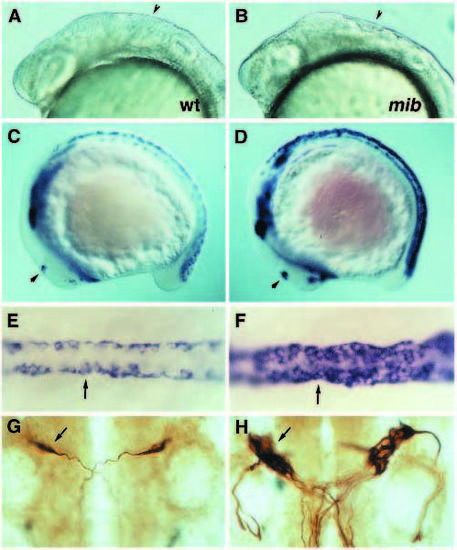
Phenotypic analysis of mind bomb (mib) mutants. (A,C,E,G) Wild type. (B,D,F,H) mind bomb (mib)m178 mutants. (A,B) Brain in wild-type (A) and mutant (B) embryos at 25-somite stage. Note the irregularities in the hindbrain (arrowhead). (C,D) Expression of islet1 in wild-type (C) and mutant (D) embryos at 13-somite stage. Note the dramatic increase of islet1-expressing cells in all regions where primary neurons are formed, including the spinal cord, epiphysis (arrow) and trigeminal ganglion. (E,F) Dorsal view of prospective Rohon-Beard cells (arrow) in dorsal spinal cord of embryos shown in C and D. (G,H) Mauthner neurons (arrow) in wild type (G) and mutant (H) embryos at 28 hpf, highlighted by 3A10 antibody. Note the abnormal projection of the most laterally located supernumerary Mauthner cell in this mutant embryo. All the other Mauthner cells project towards the midline. EXPRESSION / LABELING:
PHENOTYPE:
|

ZFIN is incorporating published figure images and captions as part of an ongoing project. Figures from some publications have not yet been curated, or are not available for display because of copyright restrictions. |
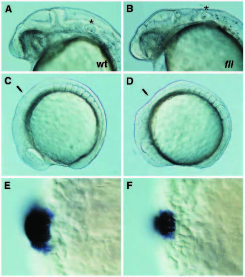
Phenotype of flachland fllm517 mutants. (A,C,E) Wild-type embryos. (B,D,F) Mutant embryos. (A,B) 31 hpf; star indicates the position of the otic vesicle and rhombomere 5. (C,D) 9-somite stage; arrow indicates the hindbrain region. (E,F) Optical cross-section through the hindbrain at the level of rhombomere 5 of 14-somite embryos stained for krox-20 and pax[zf-b] expression. Dorsal is to the left. EXPRESSION / LABELING:
PHENOTYPE:
|