- Title
-
Mutations affecting morphogenesis during gastrulation and tail formation in the zebrafish, Danio rerio
- Authors
- Hammerschmidt, M., Pelegri, F., Mullins, M.C., Kane, D.A., Brand, M., van Eeden, F.J., Furutani-Seiki, M., Granato, M., Haffter, P., Heisenberg, C.P., Jiang, Y.J., Kelsh, R.N., Odenthal, J., Warga, R.M., and Nüsslein-Volhard, C.
- Source
- Full text @ Development
|
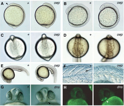
Morphological characteristics of (A-F) one-eyed pinhead (oeptz257) and (G,H) dirty nose (dnste350) mutant embryos. In each section, wild type (+) is to the left, mutant to the right. (A) Tailbud stage, lateral view: the body axis is shorter; no pillow (indicated by an arrowhead) is formed at the anterior end of the hypoblast. (B,C) 8-somite stage: (B) lateral view, the head is reduced while the tailbud seems enlarged in oep mutants. (C) dorsal view of head region. (D) 24 hours, dorsal view of head region: the anterior part of the forebrain is missing while the eye vesicles are fused in oep mutants. (E) 24 hours, lateral view: the notochord is slightly undulated, the somites are slightly expanded along the a/p axis, the yolk tube is shorter and thicker, and the tail is curled down in oep mutants. (F) 24 hours, lateral view at level of yolk tube: no floorplate (arrow) can be detected in oep mutants. (G,H) dns, 24 hours, ventral view of head region: (G) brownish cells can be observed underneath the eyes (arrowhead); (H) acridine orange staining reveals apoptotic cells in the same region (arrowhead). |
|
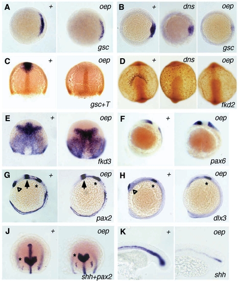
Altered gene expression in mesoderm and neuroectoderm of oep and dns mutants revealed by in situ hybridization (A-C, E-K) and immunostaining (C,D). In each section, wild type (+) is to the left, mutants to the right. (A) gsc, 40% epiboly, animal view, dorsal right. (B) gsc, shield, animal view, dorsal right. (C) gsc mRNA in blue and Ntl protein (T) in brown, 70% epiboly, dorsal view. (D) fkd2, 10-somite stage, view on head: fkd2-positive presumptive hatching gland cells are absent in oep mutants. In dns mutants, very small fkd2-positive cells can be found at the anterior border of the hypoblast rather than in adjacent regions of the yolk sac. (E) fkd3, 70% epiboly, dorsal view: fkd3 expression is missing anterior to the diencephalic-mesencephalic boundary in oep mutants. (F) pax6, 3-somite stage, lateral view: the pax6 expression domain in the presumptive diencephalon reaches the anterior border of the CNS in oep mutants. (G) pax2, 12-somite stage, lateral view: pax2 expression is normal in the otic placode (asterisk) and in the midbrain (arrow) while the expression domain in the optic stalk (triangle) is missing. (H) dlx3, 12-somite stage, lateral view: the dlx3 expression is normal in the otic placode (asterisk) but absent in the olfactory placode (triangle) in oep mutants. (J) shh and pax2, tailbud-stage, view on head region: no shh expression can be detected in the brain anterior of the pax2-positive midbrain (asterisk). The axial transcripts posterior of the midbrain are located in the notochord. (K) shh, 22 hours, lateral view on tail region: shh expression is normal in the slightly undulated notochord but absent in the ventral cells of the spinal cord. EXPRESSION / LABELING:
PHENOTYPE:
|
|
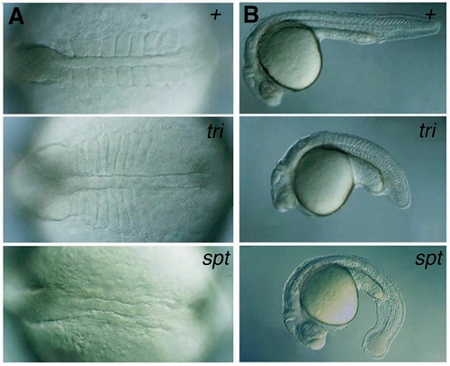
Alterations in morphology in trilobite (tritk50) and spadetail (sptb104) mutants, compared to wild type (+). (A) 8-somite stage, dorsal view: both notochord and somites are shorter and wider in tri mutants. No somites are formed in spt mutants. (B) 30 hours, lateral view: tri embryos are shorter than wild-type and spt mutant embryos. Note the accumulation of cells at the tip of the tail in spt mutants. PHENOTYPE:
|
|
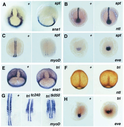
Altered gene expression in the mesoderm of spadetail (sptb104) and trilobite (tritk50) mutants compared to wild type (+), detected by in situ hybridization (A-E,G,H) and immunostaining (F). (A) sna1, 80% epiboly, dorsovegetal view. (B) ntl, 4-somite stage, dorsovegetal view. (C) myoD, 4-somite stage, dorsal view. (D) eve1, 4-somite stage, vegetal view. (E) sna1, 70% epiboly, dorsovegetal view. (F) ntl, 95% epiboly, dorsal view. (G) myoD, 10-somite stage, dorsal view on spread embryos, anterior on top. (H) eve1, 10-somite stage, vegetal view. EXPRESSION / LABELING:
PHENOTYPE:
|
|
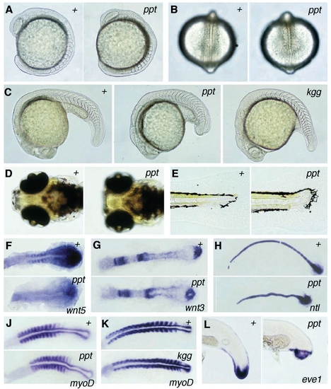
Alterations in morphology and gene expression of pipetail (pptta98, A-J,L) and kugelig (kggtl240, C,K) mutant embryos, compared to wild type (+). (A) 10-somite stage, lateral view. (B) 10- somite stage, dorsal view: in ppt mutants, the notochord is undulated and the somites are condensed along the anteroposterior axis. Somites 9 and 10 and the presomitic mesoderm show a slight lateral expansion. (C) 24-somite stage, lateral view. (D) Day 5, ventral view of head: the head skeleton forms but fails to extend beyond the eyes, leading to a slight broadening of the head in ppt mutants. (E) Day 5, lateral view of tip of tail: the notochord and the neurocoel are slightly kinked at the end of the tail; the ventral gap of the melanophore stripe is present. (F-K) Dorsal view on spread embryo, anterior left: (F) wnt5, 5-somite stage: the expression of wnt5 is significantly reduced in somites and tailbud of ppt mutants. (G) wnt3, 5-somite stage. (H) ntl, 8-somite stage. (J) myoD, 12-somite stage: all somites are condensed, the posterior somites are laterally expanded in ppt mutants. (K) myoD, 16-somite stage: the posterior somites are condensed but not laterally expanded in kgg mutants. (L) eve1, 28- somite stage, lateral view on tail. EXPRESSION / LABELING:
PHENOTYPE:
|

Unillustrated author statements PHENOTYPE:
|

