
- Title
-
Retinoic acid alters photoreceptor development in vivo
- Authors
- Hyatt, G.A., Schmitt, E.A., Fadool, J.M., and Dowling, J.E.
- Source
- Full text @ Proc. Natl. Acad. Sci. USA
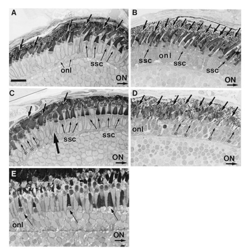
(A–D) Transverse sections of normal (A and C) and RA-treated (B and D) embryos on day 5 pf. In these sections, the optic nerve (ON) is to the right. (A) In the midperipheral region of the dorsal retina of control embryos, short single cones (ssc) with broad outer segments lie vitreal to rods and longer single cones. The short single cones are more obvious to the right, i.e., more centrally. Rod outer segments average 6 μm in length and are observed across all of this retinal region (arrows). (B) After a 3-day treatment with RA, the number of observable rods (arrows) has increased within the dorsal retina, and the short single cones (ssc) are smaller and less obvious. (C) The ventral retina in control eyes is partitioned into two compartments: a cone-dominated region (right of large arrow) adjacent to the optic nerve and a peripheral region (left of large arrow) dominated by rods (arrows). Mature short single cones (ssc) lie vitreal to a layer of longer single cones in the cone-dominated region. In the rod-dominated region, a population of miniature cones (asterisks) are seen. (D) After RA treatment beginning at day 2 pf, the cone-dominated region within the ventral retina is not observed. Rather large rod outer segments (arrows) are observed extending across the entire ventral region rather than being restricted to the periphery. These rod outer segments appear less organized than those of controls and average ≈10 μm in length, which is ≈2 μm longer than those observed in the ventral region of controls. Small cones (asterisks) resembling the miniature cones located within the ventral periphery in controls extend across the entire ventral region. (E) Transverse section of the peripheral retina from a normal retina at 21 days pf. Arrows identify maturing short single cones (from left to right). onl, Outer nuclear layer. (A–D, bar = 16 μm; E, bar = 9 μm.) |
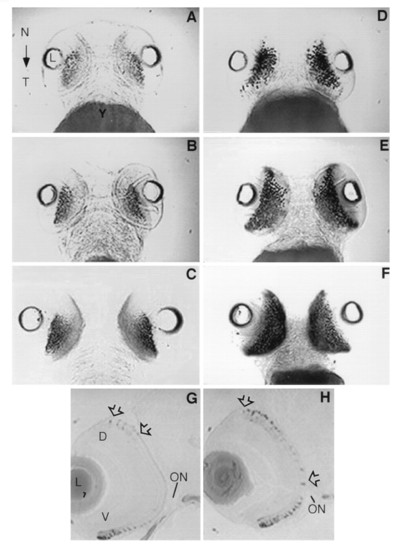
Control embryos (A–C) and RA-treated embryos (D–F) in ventral view showing the localization of rhodopsin mRNA expression by whole mount in situ hybridization. In controls, the level of rhodopsin mRNA expression increases modestly within the ventral retina during development through 3 (A), 4 (B), and 5 (C) days pf. After a 24-hr RA treatment beginning at day 2, staining within the ventral region is more robust indicating the level of rhodopsin expression has increased significantly (D). Not only is rhodopsin expression greater but it extends further into the nasal and temporal regions of the retina as compared with controls. After 2 and 3 days of RA treatment, the level of rhodopsin expression is further increased relative to the controls and extends throughout the ventral region as far as the nasal and temporal margins at days 4 (E) and 5 pf (F), respectively. The localization of rhodopsin transcripts by in situ hybridization at day 4 in transverse sections (5 μm) in control (G) and RA-treated (H) embryos. (G) On day 4, rhodopsin expression is evident within the periphery of the ventral retina (V). Weak expression is also observed near the dorsal (D) periphery (arrows). (H) After 2 days of RA treatment, rhodopsin expression extends throughout the ventral retina. Furthermore, expression is considerably more robust and extensive within the dorsal retina (arrows). ON, optic nerve; N, nasal region; T, temporal region; L, lens; Y, yolk. |
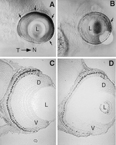
Localization of mRNA transcripts for ultraviolet opsin by whole mount in situ hybridization at day 5 pf in control embryos (A) and RA-treated embryos (B) in lateral view. (A) In controls, ultraviolet opsin expression is most robust within the nasal retina and faint expression is also observed in the temporal retina (arrows indicate regions of high expression). (B) After three days of RA treatment, ultraviolet opsin expression is significantly decreased at day 5 in RA-treated embryos throughout most of the retina. The only region of dense staining is found within the nasal retina (arrow). The localization of blue opsin transcripts by in situ hybridization at day 5 in transverse sections (5 μm) in control (C) and RA-treated (D) embryos. Blue opsin staining is generally weaker in RA-treated embryos (D). Furthermore, staining patterns of cells expressing the blue opsin appears graded in RA-treated embryos along the dorsal-ventral axis of the retina. The size of cones expressing the blue-sensitive opsin is decreased, particularly in regions ventral to the dorsal retina. D, dorsal retina; V, ventral retina; L, lens; Y, yolk. |
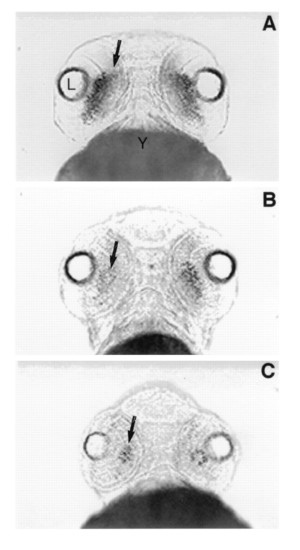
Ventral views of rhodopsin expression in whole mount in situ preparations at day 3 pf in control embryos (A) and embryos treated with the RA-synthetic competitive inhibitor citral (B and C). (A) Rhodopsin expression (arrow) extends across the ventral retina in control embryos at day 3. (B) After treatment with 3 μM citral between days 2 and 3 pf, the level of rhodopsin expression (arrow) is significantly reduced. (C) After treatment with 6 μM citral, rhodopsin expression is further reduced such that only a small number of rods (arrow) are observed within the retina at day 3 pf. L, lens; Y, yolk. |