
- Title
-
Identification of separate slow and fast muscle precursor cells in vivo, prior to somite formation
- Authors
- Devoto, S.H., Melancon, E., Eisen, J.S., and Westerfield, M.
- Source
- Full text @ Development
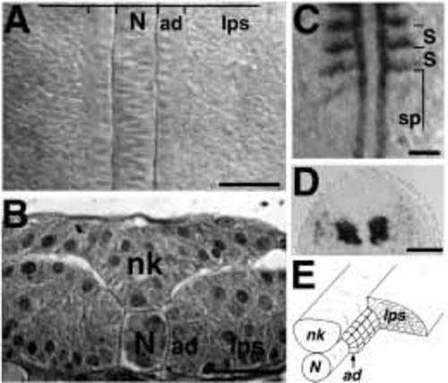
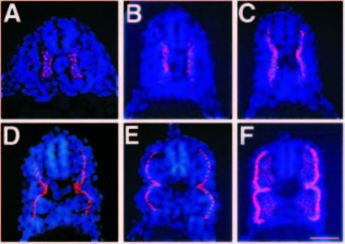
Adaxial and lateral presomitic cells can be distinguished in the segmental plate. (A) Dorsal view of the segmental plate of an approximately 13 h (8 somites have formed) live embryo. The cuboidal adaxial cells (ad) on either side of the notochord (N) can be distinguished from the irregularly shaped lateral presomitic cells (lps). (B) Transverse semithin section of the segmental plate of a 12 h (6 somites) embryo. The adaxial cells (ad) are large cells adjacent to the notochord (N). Lateral presomitic cells (lps) are smaller, more irregularly shaped and not contacting the notochord. nk, neural keel. (C) Dorsal view of a 13 h (8 somites) embryo labeled by wholemount RNA in situ hybridization for myoD. In the segmental plate (sp), only adaxial cells abundantly express myoD; as somites (S) form, other somitic cells also express myoD. Horizontal lines mark somite borders. (D) Transverse section through the most rostral portion of the segmental plate of an approximately 15 h (12 somites) embryo labeled by whole-mount in situ hybridization for myoD. Adaxial cells express very abundant levels of myoD. Some of the lateral presomitic cells are beginning to express low levels of myoD. (E) Schematic drawing of the segmental plate. The adaxial cells (ad) are arranged as a sheet between the notochord (N) and the lateral presomitic cells (lps); the neural keel (nk) is also shown. Approximately 20 adaxial cells contribute to each somite. In dorsal views, anterior is to the top of the figure; in transverse sections, dorsal is up. Scale bars: A,B, 25 μm; C, D, 50 μm. EXPRESSION / LABELING:
|
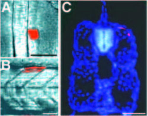
Adaxial cells become the most superficial muscle cells in the somite. (A) Dorsal view of the segmental plate of an approximately 15 h (12 somites) live embryo, after injection of two adaxial cells with lysinated rhodamine dextran. (B) Side view of the same embryo at about 40 h. Both of the injected cells developed into dorsal muscle cells in somite 15 (C) Transverse section of the same embryo, counter stained with Hoechst 33258 to show cell nuclei (blue). The two injected adaxial cells (red) are located superficially. In a series of similar experiments, 72 out of 73 injected adaxial cells became superficial muscle fibers (the single exception was a dorsal cell that may have been adjacent to the neural keel instead of the notochord, and hence misidentified as an adaxial cell at the time of injection). In side views, anterior is to the left; in transverse sections, dorsal is up. Scale bars, 50 μm. |
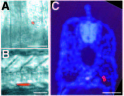
Lateral presomitic cells become deep muscle cells. (A) Dorsal view of the segmental plate of an approximately 15 h embryo (12 somites), after injection of a lateral presomitic cell with lysinated rhodamine dextran. (B) Side view of the same embryo at about 40 h. The lateral presomitic cell developed into two ventral muscle cells located in somite 16. (C) Transverse section of the same embryo, counter stained with Hoechst 33258 to show cell nuclei. Both cells are deep muscle fibers. In a series of similar experiments, 25 out of 25 injected lateral presomitic cells became deep muscle fibers. In side views, anterior is to the left; in dorsal views, anterior is up; in transverse sections, dorsal is up. Scale bars, 50 μm. |
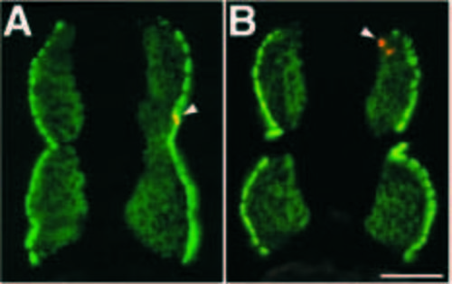
F59 intensely labels a monolayer of superficial muscle cells derived from adaxial cells and weakly labels deep muscle cells derived from lateral presomitic cells. Adaxial (A) or lateral presomitic (B) cells were injected at about 14 h with vital dye. At 40 h, embryos were fixed and sections stained with F59. (A) The injected adaxial cell (arrowhead, yellow due to double fluorescence of red and green) became a superficial muscle cell strongly labeled by F59 (green). (B) The injected lateral presomitic cells (arrowhead, red) became deep muscle cells only weakly labeled by F59 (green). In a series of similar experiments, 6 out of 6 injected adaxial cells became part of the superficial layer of strongly F59 reactive muscle fibers; 12 out of 12 injected lateral presomitic cells differentiated into the deeper, weakly F59 reactive muscle fibers. We have also used the monoclonal antibody zn-5, which labels the same population of superficial muscle cells that are strongly labeled by F59 at this time (data not shown). In a series of similar experiments, 18 out of 19 injected adaxial cells differentiated into one of the superficial monolayer of muscle fibers positive for zn-5 (the single exception was the same cell mentioned in the legend to Fig. 2, that was dorsal and may have been adjacent to the neural keel instead of the notochord). In these transverse sections, dorsal is up. Scale bar, 50 μm. EXPRESSION / LABELING:
|
Adaxial cells migrate radially away from the notochord, between other somitic cells. In the caudal trunk, migration occurs over approximately 5 hours. Transverse sections through the caudal trunk (somites 14-17) were immunolabeled with F59 to mark adaxial cells and counter stained with Hoechst to reveal the nuclei (A-F). (A) 17 h (16 somites) embryo. F59 immunoreactivity is present only in adaxial cells, the labeling is perinuclear. (B) 18.5 h (19 somites) embryo. The adaxial cells have begun to move dorsally and ventrally; this is approximately the time that these cells are elongating in the anterior posterior dimension. (C) 20.5 h (23 somites) embryo. The adaxial cell population now extends almost the full dorso ventral extent of the myotome. (D) 21.5 h (25 somites) embryo. Radial migration of the adaxial cells has begun. (E) 23 h (28 somites) embryo. Most of the adaxial cells have reached the lateral surface. (F) 24 h embryo. The adaxial cells are now lateral. A subset of adaxial cells remains apposed to the notochord, generating an hourglass shape to the layer of superficial muscle cells. The deep cells of the myotome are now beginning to express the F59 epitope weakly. In these transverse sections, dorsal is up. Scale bar, 50 μm. EXPRESSION / LABELING:
|
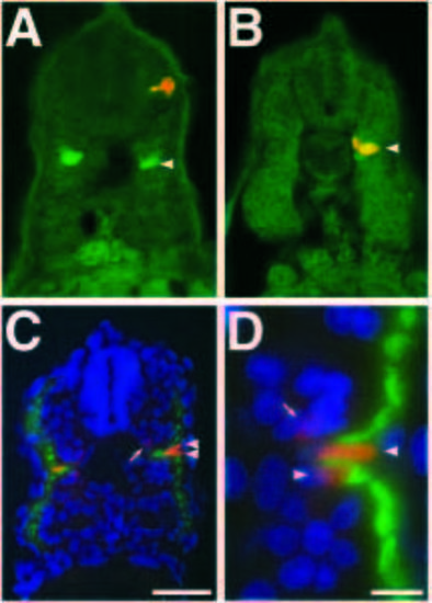
Muscle pioneer cells are a subset of adaxial cells. (A,B) Transverse sections of ~22 h embryos in each of which an adaxial cell had been injected as in Fig. 2. Sections were immunostained with the 4D9 Engrailed antibody, which labels the muscle pioneers (green). One out of 7 injected adaxial cells became Engrailed-labeled muscle pioneers. (A) An adaxial cell (red) that became a superficial muscle cell that is not a muscle pioneer (green, arrowhead). (B) An adaxial cell (arrowhead, yellow due to double labeling in green and red fluorescence) that differentiated into an Engrailed-expressing muscle pioneer. (C) Transverse section through a 24 h embryo immunolabeled with the S58 myosin antibody (green) to mark the superficial muscle cells derived from adaxial cells and the 4D9 engrailed antibody (red) to label the muscle pioneers. The Engrailed-labeled muscle pioneers (arrowheads, bright red nuclei) are a subset of the superficial muscle cells. Other somite cells near the muscle pioneers also weakly express Engrailed (arrow, Hatta et al., 1991a), these cells are unlabeled by the S58 antibody and are not part of the superficial muscle cell population derived from adaxial cells. (D) Higher magnification view of the region of the myotome containing muscle pioneers. The two cells that express Engrailed at high levels (arrowheads, bright red nuclei), are also S58 positive (green). Another somite cell that weakly expresses Engrailed, not a muscle pioneer, does not express S58 and is not part of the adaxialderived superficial muscle cells (arrow, nucleus light red). In these transverse sections, dorsal is up. Scale bars: A-C, 50 μm, D, 10 μm. EXPRESSION / LABELING:
|
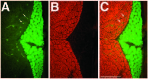
Slow and fast muscle fibers can be unambiguously distinguished by immunolabeling. A transverse section of a 32 d zebrafish was doubly immunolabeled with the S58 antibody and the 12/101 antibody (A-C). (A) S58 (green) recognizes only the slow muscle fibers. Some non-specific fluorescence is present within blood vessels in all muscle regions (e.g. arrows). (B) 12/101 (red) recognizes fast and intermediate, but not slow muscle fibers. (C) 12/101 (red) and S58 (green) label mutually exclusive muscle fibers. In these transverse sections, dorsal is up. Scale bar, 100 μm. EXPRESSION / LABELING:
|
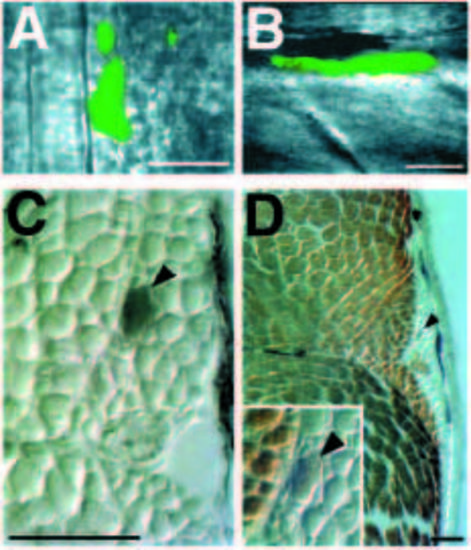
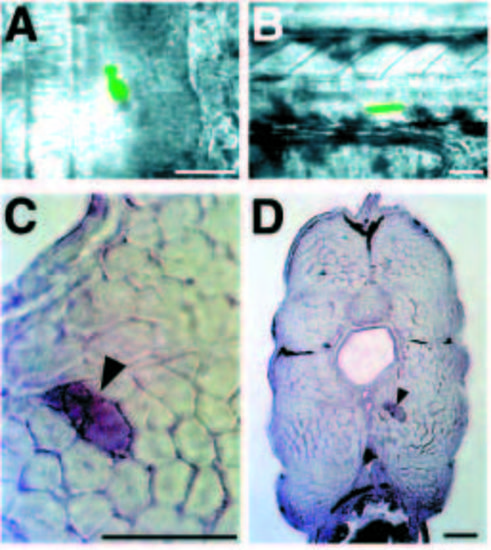
Adaxial cells become slow muscle fibers. (A) Dorsal view of the segmental plate of an approximately 12 h embryo (6 somites), after injection of adaxial cells with lysinated fluorescein dextran. Two or three adaxial cells were injected in this embryo, acellular fluorescent debris is present lateral to the injected adaxial cells. (B) Side view of the same embryo at 8 days. Only one of the injected adaxial cells survived. It developed into a muscle cell located in somite 11. The hexagonal background pattern in this image is due to the video camera. (C) Transverse section of the same embryo at 20 d. The section has been labeled with an antibody to fluorescein, thus the injected cell is black (arrowhead). (D) The same section after labeling with the 12/101 antibody. The adaxial cell developed into a small diameter muscle fiber located in the slow muscle region, not labeled by 12/101 (arrowhead). Inset: higher magnification (as in C) view of the injected cell (arrowhead). In a series of similar experiments, 4 out of 4 injected adaxial cells became slow muscle fibers, as determined by antibody staining, position and morphology. In dorsal views, anterior is up; in side views, anterior is to the left; in transverse sections, dorsal is up. Scale bars, 50 μm. |
Lateral presomitic cells become fast muscle fibers. (A) Dorsal view of the segmental plate of an approximately 15.5 h embryo (13 somites), after injection of lateral presomitic cells with lysinated fluorescein dextran. (B) Side view of the same embryo at about 38 h of development. One of the lateral presomitic cells developed into a ventral muscle fiber located in somite 15. (C) Transverse section of the same embryo at 15 d. The injected lateral presomitic cell developed into a large diameter muscle fiber (arrowhead) located deep in the muscle. (D) Lower magnification view of the injected cell (arrowhead). The muscle fiber is within the fast muscle region. In a series of similar experiments, 3 out of 3 injected lateral presomitic cells became fast muscle fibers. In dorsal views, anterior is up; in side views, anterior is to the left; in transverse sections, dorsal is up. Scale bars, 50 μm. |