
- Title
-
Expression of the zebrafish paired box gene pax[zf-b] during early neurogenesis
- Authors
- Krauss, S., Johansen, T., Korzh, V., and Fjose, A.
- Source
- Full text @ Development
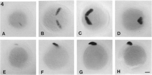
Localization of pax[zf-b] transcripts by in situ hybridization on a series of whole-mount embryos of early developmental stages between 9 and 10h. Lateral views (E-H) and dorsal views (A-D) are shown. The embryos are oriented with the anterior end to the right. Bar, 100 μm. EXPRESSION / LABELING:
|
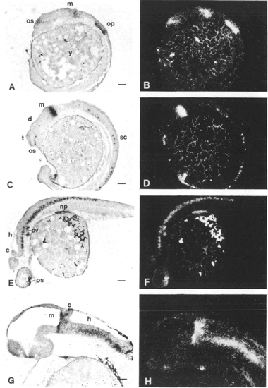
Localization of pax[zf-b] transcripts by in situ hybridization in tissue sections of zebrafish embryos at different developmental stages. Sagittal sections are shown for embryos after 12 h (A,B), 18 h (C,D), 24 h (E,F) and 36 h (G,H) of development. Bright-field and dark-field images of each section are shown side by side. The embryos are oriented with their anterior end to the left. Abbreviations: c, cerebellum; d, diencephalon; h, hindbrain; m, midbrain; np, nephritic primordium; op, otic placode; os, optic stalk; ov, otic vesicle; t, telencephalon; y, yolk. Bars, 50 μm. EXPRESSION / LABELING:
|
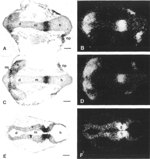
Distribution of pax[zf-b] transcripts in horizontal sections of embryos at 12 h (A,B), 18 h (C,D) and 24 h (E,F) of development. The embryos are oriented as in Fig. 3. Abbreviations: d, diencephalon; e, eye; h, hindbrain; m, midbrain; op, otic placode; os, optic stalk. Bars: 50 μm. EXPRESSION / LABELING:
|
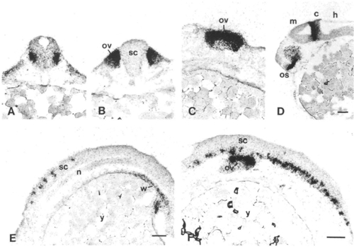
Details of the pax[zf-b] expression. (A) Cross section at the hindbrain-spinal cord junction of a 24h embryo. (B) Cross section and (C) sagittal section of the region covering the otic vesicle at 24h of development. (D) Sagittal section of a 24 h embryo demonstrating the pax[zf-b] in the optic stalk and the posterior midbrain. (E,F) Close up of the spinal cord at 18 h and 24h of development. Abbreviations: c, cerebellum; h, hindbrain; m, midbrain; n, notochord; os, optic stalk; ov, otic vesicle; sc, spinal cord; w, Wolffian duct; y, yolk. Bars: 50 μm. EXPRESSION / LABELING:
|
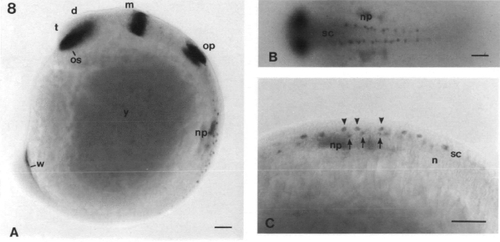
In situ hybridization of pax[zf-b] on whole-mount embryos at 15 h of development. The embryo is oriented as in Fig. 3. (A) Lateral view of the embryo. (B) Dorsal view of the spinal cord of the embryo. (C) Close up of the spinal cord seen from lateral. Dorsal is to the top, the two rows of cells described in Discussion are marked with arrows and arrowheads, respectively. Abbreviations: d, diencephalon; e, eye; h, hindbrain; m, midbrain; np, nephritic primordium; os, optic stalk; ov, otic vesicle; sc, spinal cord; t, telencephalon; w, Wolffian duct; y, yolk. Bars: 50μm. EXPRESSION / LABELING:
|