
- Title
-
Connectional topography in the zebrafish olfactory system: random positions but regular spacing of sensory neurons projecting to an individual glomerulus
- Authors
- Baier, H., Rotter, S., and Korsching, S.
- Source
- Full text @ Proc. Natl. Acad. Sci. USA
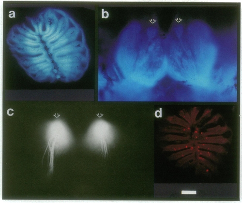
Backtracing of olfactory sensory neurons projecting into a single glomerulus. (a) Olfactory epithelium, 15 hr after CMAC injection. Cells in the central part of the organ (the sensory area) are predominantly stained. (b) Pair of olfactory bulbs, immediately before DiI injection. CMAC-labeled glomeruli are easily recognized. The vpGs are indicated by arrows. (c) Pair of olfactory bulbs, one week after DiI injection into the vpGs (arrows). Although the dye label has broadened by diffusion, labeled axons emerge only from the initial injection site. (d) Olfactory epithelium, 2 weeks after DiI injection into the vpG, displaying individual retrogradely labeled neurons. For a-d, posterior is to the top, and anterior is bottom. (Bar = 200 μm.) |