
- Title
-
Affinity purification of in vivo assembled whirlin-associated protein complexes from the zebrafish retina
- Authors
- Schellens, R.T.W., Slijkerman, R.W.N., Hetterschijt, L., Peters, T., Broekman, S., Clemént, A., Westerfield, M., Phillips, J.B., Boldt, K., Kremer, H., De Vrieze, E., Van Wijk, E.
- Source
- Full text @ J. Proteomics
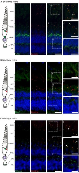
Fig. 1. SF-Whrna is present at the connecting cilium, in the inner segment, and at the synapse of the adult zebrafish photoreceptor. (A) Strep/Flag-tagged Whrna (green signal) is detected at the base of the connecting cilium, partially overlapping with centrin (red signal), and in the photoreceptor inner segments localized in the outer nuclear layer (ONL). In addition, signal is observed in the outer plexiform layer (OPL), where the photoreceptor synaptic regions are localized. (B) The retinas of wild-type zebrafish were used as negative control. No anti-FLAG signal could be detected. (C) Immunohistochemical analysis of wild-type zebrafish retinas using antibodies directed against Whrna. Whrna (green signal) is detected at the base of the connecting cilium, where it partially colocalizes with the connecting cilium marker centrin (red signal), and in the OPL, where the photoreceptor synaptic regions are localized. Co-localisation of Whrna and centrin is indicated by arrowheads. ROS = rod outer segment, RPE = retinal pigment epithelium, COS = cone outer segment, ONL = outer nuclear layer, OPL = outer plexiform layer, INL = inner nuclear layer. Scalebars represent 20 μm in the overview images and 10 μm in the higher magnification images. (For interpretation of the references to colour in this figure legend, the reader is referred to the web version of this article.) EXPRESSION / LABELING:
|
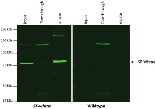
Fig. 2. Western blot analysis of FLAG-tagged proteins upon anti-FLAG affinity purification of wild-type and SF-whrna zebrafish retinal extracts. SF-Whrna (83 kDa, indicated with an arrow) was abundantly detected in the SF-whrna retinal extract (input) and in the SF-whrna eluate after affinity purification, but little was detected in the SF-whrna flow-through upon washing. In the wild-type samples, SF-Whrna was not detected. |
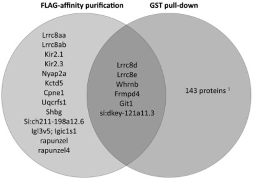
Fig. 3. Overview of Whrna-associated proteins retrieved from anti-FLAG affinity purification and/or GST pull-down assays. Proteins that have been identified as candidate Whrna interaction partners, arranged by the purification method (i.e. anti-FLAG affinity purification or GST pull-down assays). 1 Proteins retrieved by GST pull-down assays are listed in Supplemental Table 3. |
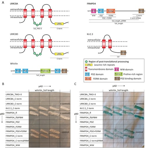
Fig. 4. Human whirlin binds directly to FRMPD4 and Kir2.3, but not to LRRC8A and LRRC8D. (A) Graphical representation of the predicted protein domain architecture of human LRRC8A, LRRC8D, whirlin, FRMPD4 and Kir2.3 (http://smart.embl-heidelberg.de). Peptides that were used in yeast two-hybrid experiments are underlined. (B) Yeast two-hybrid reporter gene activation upon binding of whirlin to candidate interaction partners. The panel shows selective yeast growth after the most stringent amino acid restriction (-WLHA). Plasmids encoding fragments of candidate interaction partners fused to a DNA binding domain (pBD) were co-expressed in yeast with a plasmid encoding full length whirlin fused to an activator domain (pAD). All combinations of peptides, except for controls, were analyzed in triplicate. (C) Analysis of β-galactosidase reporter gene activation in yeast grown on the selective medium as shown in (B). The blue colour results from the hydrolysis of the X-β-gal substrate by β-galactosidase and is indicative of an interaction between the two protein peptides. “+ control 1” and “+ control 2” are the positive controls (pAD whirlin full length with pBD usherin c-term) [11] and “- control” is the negative control. (For interpretation of the references to colour in this figure legend, the reader is referred to the web version of this article.) |
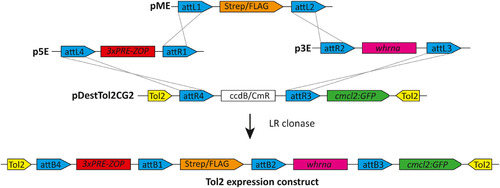
Generation of the pDestTol2CG2_3×PRE-ZOP:SF-whrna expression construct. A 3× photoreceptor regulatory element (PRE)-containing zebrafish rod opsin promoter element (ZOP), a double Strep and single FLAG-tag (Strep/FLAG) and whrna (ENSDART00000111659.4) were cloned into the pDestTol2CG2 destination vector. This resulted in the pDestTol2CG2_3xPRE-ZOP:SF-whrna expression construct, which further contains the heart-specific cmcl2 promoter driving the expression of EGFP.
Construct:
Tg(rho:SFtag-whrna,myl7:EGFP)
|
Reprinted from Journal of proteomics, 266, Schellens, R.T.W., Slijkerman, R.W.N., Hetterschijt, L., Peters, T., Broekman, S., Clemént, A., Westerfield, M., Phillips, J.B., Boldt, K., Kremer, H., De Vrieze, E., Van Wijk, E., Affinity purification of in vivo assembled whirlin-associated protein complexes from the zebrafish retina, 104666, Copyright (2022) with permission from Elsevier. Full text @ J. Proteomics