FIGURE SUMMARY
Title

A Zebrafish Forward Genetic Screen Identifies an Indispensible Threonine Residue in the Kinase Domain of PRKD2

Authors
Giardoglou, P., Bournele, D., Park, M., Kanoni, S., Dedoussis, G.V., Steinberg, S.F., Deloukas, P., Beis, D.
Source
Full text @ Biol. Open

. s411 carry a mutation in the prkd2 gene. (A,B) Bright field image analysis of a wild-type zebrafish embryo compared to an s411 mutant embryo at 72 hpf. Heterozygous adults that carry the s411 recessive mutation give 25% offspring mutant embryos exhibiting heart edema, inadequate blood circulation leading to complete outflow tract stenosis and blood regurgitation by 72 hpf. Scale bars: 500 μm. (C) Bright-field image analysis prkd2-targeted morpholino-injected embryo at 72 hpf. MO-prkd2-injected embryos resemble the s411 phenotype possessing the same features as s411 mutants (three replicates, n>50). Scale bar: 200 μm. (D) Bulked segregant analysis and further genetic mapping positioned the s411 mutation on chromosome 15. Recombination analysis on 865 embryos, positioned the mutation initially between the markers, z6895 and z51478 and further fine-mapping positioned the mutation between markers we developed in the overlapping BACs: CU062627 and BX924136. (E) The s411 embryos carry an A to G mutation that translates to a Threonine (T) to Alanine (A) amino acid change. (F) Blast analysis showed that the threonine T757 (T714 in humans) is a conserved amino acid at a highly conserved region of the C-terminus PRKD2 kinase, between several species. (G) Schematic representation of zebrafish Prkd2 kinase. It consists of 923 amino acids and the main domains of the enzyme are indicating: two cysteine-rich motif domains (cys1 and cys2), a pleckstrin homology domain (PH), and the C-terminal catalytic domain where the PKC-phosphorylation sites, Ser749 and Ser753 reside. It is also highlighted the position of s411 mutation (A to G), a previously identified zebrafish mutation with a similar phenotype (Y849) and the position of premature stop codon after MO injection resulting to defective splicing.

The T757A substitution disrupts PRKD2 kinase activity. Lysates from HEK293 cells that heterologously overexpress wild-type or mutant forms of PKD1 and PKD2 were subjected to in vitro immuno-complex kinase assays (IVKAs) in the absence or presence of PS/PMA and immunoblot analysis was used to track PKD C-tail autophosphorylation as well as PKD1 phosphorylation of recombinant CREB (added as a heterologous substrate). All results were replicated in three separate experiments.

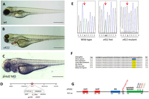
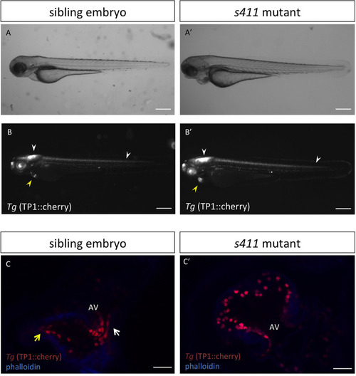
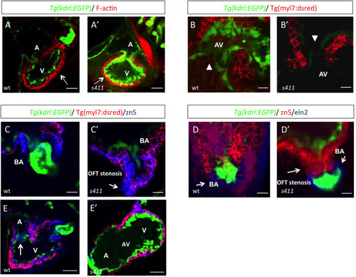
s411 embryos show aortic valve stenosis. (see also Movies 2 and 3). Confocal images of the heart (A,A′,E,E′), the AV canal (B,B′), the outflow track (C,C′) and the bulbus arteriosus (D,D′) at 72 hpf. Tg(kdrl::EGFP) (green) embryos were stained for F-actin (red) (A,A′) or zn5 (pseudo-colored red) and eln2 (pseudo-colored blue) (D,D′). Tg(kdrl::EGFP) (green)/Tg(myl7:DsRed) embryos were stained for zn5 (pseudo-colored blue) (C,C′,E,E′). (A,A′) In s411 embryos, the myocardium appears thinner compared to the wild-type embryos. Arrows indicate the myocardial wall. (B,B′) Endocardial cells residing at AV canal extend in two layers at wild-type embryos whereas the s411 mutants obtain a single-layer of AV endocardial cells. Arrowheads indicate the endocardial cells of AV. (C,C′) The endocardial cells of outflow track are in tight contact in s411 mutant embryos. This structural feature leads to stenosis formation. Arrow indicates the junction between the neighboring endocardial cells. (D,D′) Bulbus arteriosus consists of endocardial cells surrounded by a layer of smooth muscle cells. s411 mutants have a blocked bulbus arteriosus. Arrows show the elastin-positive cells and the stenosis of mutant embryos. (E,E′) In s411 embryos, the endocardial monolayer of AV cells does not express zn5 compared to the two-layer zn5+ AV endocardial cells in wild-type embryos, which indicates the malformation of atrioventricular cardiac valve during development. A, atrium; V, ventricle; AV, atrioventricular, BA, bulbus arteriosus. n=10 in each of three independent experiments. Scale bars: 50 μm for A,A′, E,E′ and 20 μm for B–D′.

Atrioventricular canal markers remain expressed throughout the heart in s411 mutant embryos. Ectopic expression of atrioventricular canal (AV) markers in s411 mutant embryos as shown by wholemount in situ hybridization. notch1b antisense probe in lateral (A,B) and dorsal (A′,B′) view of wild-type embryos (A,B) and s411 mutants, respectively (A′,B′), at 72 hpf. klf2a in wild-type (C) and s411 mutants (C′) and bmp4 in wild-type (D) and s411 mutants (D′) at 72 hpf. All three AV differentiation markers show restricted expression pattern in the AV and outflow tract of wild-type embryos, while they remain expressed throughout the heart in s411 mutants. n=10 in each of three independent experiments. Scale bars: 150 μm.

Ectopic activation of endocardial Notch signaling in s411 embryos carrying the T757A PRKD2 mutation. Bright field (A,A′) and fluorescence (B,B′) analysis of s411 sibling and mutant embryos carrying the Tg(Tp1:mCherry) (pseudo-colored grey). Scale bars: 500 μm. Confocal analysis of 150 μm cardiac slices of s411 sibling and mutant embryos carrying the Tg(Tp1:mCherry) co-stained with 633-phalloidin (blue) (C,C′); Scale bar: 25 μm. Notch signaling is active in several tissues and organs throughout the embryo and restricted to the AV canal and OFT of wild-type embryos at 72 hpf (B,C). In s411 mutants, Notch signaling appears unaffected in the embryo (B′, white arrowheads) but remains active throughout the ventricular endocardium (B′, yellow arrowheads, C′). White and yellow arrows (C) indicate the Notch-positive cells at AV canal and OFT, respectively. AV, atrioventricular; OFT, outflow tract. n=10 in each of three independent experiments.

Disruption of calcineurin/NFATc signaling and blocking PRKD activity exhibits a similar phenotype to s411 mutant embryos. Incubation of wild type (A) with 10 μg ml−1 cyclosporine A (CsA) results to a s411 phenotype. s411 heterozygous (B) embryos treated with cyclosporine A, at 2 μg ml−1, a sublethal dose of CsA show phenotype while there is no effect in wild-type embryos (C) when treated at this dose. (D) Treatment of wild-type embryos with a protein kinase D family inAhibitor, 2,3,4,5 -Tetrahydro-7-hydroxy- 1H-benzofuro[2,3-c]azepin-1-one between 30–72 hpf phenocopies s411 mutants showing that this is the critical window of prkd2 activity in the heart. n=20 in each of three independent experiments. Scale bars: 100 μm.

PRKD2 association with TBX5a regulation. Multiple predicted transcription factor Tbx5a binding sites in the promoter region of zebrafish Prkd2 via the ConTraV3 web server (A) and the possible DNA binding motif (B). Brightfield image analysis and quantification of 72 hpf tbx5a-morpholino injected s411 siblings, tbx5a-morpholino injected wild-type and control wild-type embryos (C). tbx5-morpholino injected s411 mutant embryos exhibit an earlier more severe phenotype reminiscent to the tbx5 morphants and mutants. Scale bars: 100 μm. As measured via rt-qPCR, prkd2 expression levels in tbx5 morphants are reduced compared to uninjected siblings at 56 hpf, normalized both to act2b and elf1a as reference genes (D). n=6 independent replicates, 15 larvae per sample, Student's t-test (two-tailed distribution, paired), ** significantly different P-value <0.01, error bars +s.e.m.

Acknowledgments
This image is the copyrighted work of the attributed author or publisher, and ZFIN has permission only to display this image to its users. Additional permissions should be obtained from the applicable author or publisher of the image. Full text @ Biol. Open