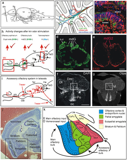
Teleostean and mammalian amygdala and accessory olfactory system. a Lateral view of adult zebrafish (Danio rerio) brain with indication of section levels shown for olfactory bulb (d/e) and telencephalon (f/g). b Schema shows the three main classes of teleostean/zebrafish olfactory sensory neurons (OSNs). Crypt cells, as all other OSN types, are widely distributed over the entire olfactory epithelium (modified after Kress et al. 2015). c Double-label immunohistochemistry for calcium-binding proteins in adult zebrafish olfactory epithelium demonstrates that calretinin (as also calbindin1 and parvalbumin, both not shown here) is absent in crypt cells which are solely characterized by S100 (modified after Kress et al. 2015). d, e Transverse sections through adult zebrafish olfactory bulb show parvalbumin (d) and S100 (e) immunostained fibers (modified after Kress et al. 2015; see there for details). Lower key green letters in d designate groups of olfactory bulb glomeruli, white letters designate calretinin/calbindin1-free glomerular groups, e.g., the mediodorsal bulbar glomeruli. Apparent parvalbumin fibers in mdG2 originate from a subpopulation of microvillous OSNs, not from crypt cells. e S100 fibers from crypt cells converge into one glomerulus, i.e., mdG2. Red elements at bulbar periphery are glial. f, g Neuroanatomical identification of the intermediate nucleus of ventral telencephalon (Vi) as the homolog of the medial amygdala using nuclear stain DAPI (f) and immunohistochemistry for Otpa (g) (modified after Biechl et al. 2017). h, i Schematics of primary and secondary olfactory pathways in adult zebrafish (modified after Biechl et al. 2017). h Neuronal activity as quantified with pERK at three successive synaptic levels from peripheral OSNs in the olfactory epithelium to two consecutive central nervous targets, including the olfactory bulb glomerulus mdG2 (immunohistochemically identified with S100 antibody because the projections of S100 immunopositive crypt cells terminate there; see text and e) and the intermediate nucleus of the telencephalon Vi (immunohistochemically identified with Otpa antibody for many of its cell bodies, see text and g) after kin odor stimulation of kin-imprinted and non-imprinted larvae. The counted pERK-positive cells were located in the olfactory epithelium, around the mdG2 and within Vi. Red tickmarks indicate statistically significant changes in activated cell numbers seen at each level (see Biechl et al. 2017 and text for details). Adult secondary olfactory projections of mediodorsal bulbar area are indicated with solid black lines (targets shared with projections of entire olfactory bulb) and dashed black lines (targets specifically attributed to mediodorsal bulbar area; see literature below). i Projections of adult zebrafish mediodorsal olfactory bulb area (including mdG2) shown after lipophilic tracing substance DiI injection (Biechl et al. 2017). Additional tracer injections into tuberal hypothalamus (TH) demonstrate furthermore a teleostean accessory olfactory pathway via subpallial amygdalar nuclei Vp/Vi. Olfactory bulb projections shown as dashed lines to telencephalic targets are selective for mediodorsal olfactory bulb (our data are shown in red lines, confirmed and extended by additional data from Ahuja et al. 2013; Kress et al. 2015; Sato et al. 2005; Braubach et al. 2012; Miyasaka et al. 2009; Gayoso et al. 2012; Turner et al. 2016). j Transverse Nissl-stained frontal section through left mouse forebrain (Mus musculus, section by courtesy of Dr. Alex Kaiser) with major telencephalic and diencephalic regions indicated. Frame indicates region shown schematically in k. k Mouse amygdala and olfactory (piriform) cortex. Blue: olfactory (piriform) cortex and endopiriform nuclei. Green: pallial amygdala (lateral, basolateral, basomedial, and cortical amygdala). Red: subpallial amygdala (amygdalo-striatal transition area, central, and medial amygdala, intra-amygdalar bed nucleus of stria terminalis) (redrawn and simplified after Martínez-García et al. 2012). Main olfactory epithelium input reaches via main olfactory bulb the piriform cortex and most (pallial) cortical amygdalar nuclei (but not the large posteromedial nucleus, not shown in k) as well as the dorsal part of the (subpallial) medial amygdala. Vomeronasal organ input reaches via accessory olfactory bulb the pallial part of the anterior amygdala (not shown), the entire medial amygdala and part of the cortical amygdala (its anterior nucleus, CoA, and, in particular, its large posteromedial nucleus (not shown). No vomeronasal input reaches the posterolateral cortical amygdalar nucleus, CoPL. Abbreviations: AStr, Amygdalo-striatal transition area; BL, Basolateral (or basal) amygdala; BM, Basomedial (or accessory basal) amygdala; BSTa, Intra-amygdalar part of bed nucleus of the stria terminalis; CaPu, Caudate-putamen (striatum); CC, Crista cerebellaris; CCe, Corpus cerebelli; CeA, Central amygdala; CilCells, Ciliated cells (olfactory sensory neurons); CoA, Anterior part of cortical amygdala; CoPL, Posterolateral part of cortical amygdala; Cr, Crypt cells; DEn, Dorsal endopiriform nucleus; Dl, Lateral zone of dorsal telencephalic area; Dm, Medial zone of dorsal telencephalic area; Dp, Posterior zone of dorsal telencephalic area; EG, Eminentia granularis; GlPa, Globus pallidus; Ha, Habenula; Had, Dorsal part of Ha; Hav, Ventral part of Ha; L, Lateral amygdala; LI, Hypothalamic lobus inferior; LL, Lateral line nerves; LVe, Lateral ventricle; mdG, Mediodorsal group of olfactory bulb glomeruli; mdG2, Mediodorsal olfactory bulb glomerulus 2; MeA, Medial amygdala; MO, Medulla oblongata; MS, Medulla spinalis; oc, Optic chiasma; OB, Olfactory bulb; oc, Optic chiasma; OE, Olfactory epithelium; Olf (pir) cortex, Olfactory (piriform) cortex; oln, Olfactory nerve; Pit, Pituitary; Po, Preoptic region; SC, Spinal cord; Tel, Telencephalon; TeO, Optic tectum; TH, Tuberal hypothalamus; TLa, Torus lateralis; Vd, Vi, Vp, Vs, Vv, Dorsal, intermediate, postcommissural, supracommissural, ventral nucleus of ventral telencephalic area; VEn Ventral endopiriform nucleus; VE, ventral entopeduncular nucleus; VLo/LX, Vagal lobe. Cranial nerves: I Olfactory nerve, II Optic nerve, IV Trochlear nerve, VII Facial nerve, VIII Octaval nerve, X Vagal nerve
|