FIGURE SUMMARY
Title

A triple-drug nanotherapy to target breast cancer cells, cancer stem cells, and tumor vasculature

Authors
El-Sahli, S., Hua, K., Sulaiman, A., Chambers, J., Li, L., Farah, E., McGarry, S., Liu, D., Zheng, P., Lee, S.H., Cui, J., Ekker, M., Côté, M., Alain, T., Li, X., D'Costa, V.M., Wang, L., Gadde, S.
Source
Full text @ Cell Death Dis.

Combination of verteporfin-NP, paclitaxel-NP, and/or combretastatin-NP suppressed both bulk tumor cells and cancer stem cells.

A The viability of TNBC MDA-MB-231 cells was determined by MTT assay after 120-hr treatment with E-NP (empty-nanoparticle), P-NP (paclitaxel-NP, 5 nM), V-NP (verteporfin-NP, 250 nM), C-NP (combretastatin-NP, 5 nM), or different combinations. B Tumorsphere (TS) formation, an in vitro assay indicating cancer stem cells (CSCs) after different treatments with drug-NPs. MDA-MB-231 cells overexpressing E-cadherin were grown in low attachment and serum-free conditions, and treated for 120 hrs as described in A, followed by MTT viability assay. C Representative photographs of TS shown in B. Data represent means ± SD, *P < 0.05, **P < 0.01, ****P < 0.001.

Verteporfin-NP more effectively inhibits the migration of TNBC cells and fully suppresses YAP target genes upregulated by paclitaxel-NP and/or combretastatin-NP.

A TNBC MDA-MB-231 cells were grown to confluence, treated with mitomycin to inhibit cell proliferation, and then scratched and quantified for migration after incubation with E (empty-NP), 5 nM P (paclitaxel-NP), 250 nM V (verteporfin-NP), 5 nM C (combretastatin-NP), or different combinations for 48 hrs. B Representative images of A. C, D RT-qPCR analysis of changes of YAP target gene in MDA-MB-231 cells after 24 -hr of treatment with E (empty-NP), 25 nM P (paclitaxel-NP), 1.25 µM V (verteporfin-NP), 25 nM C (combretastatin-NP), and different combinations. Data represent means ± SEM, *P < 0.05, **P < 0.01, ****P < 0.0001.

Verteporfin in combination with combretastatin more effectively inhibits vasculature in transgenic zebrafish (Tg, fli:eGFP) embryos.

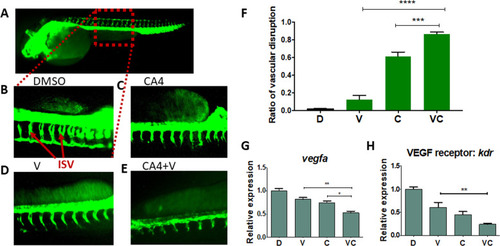
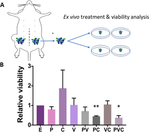
The zebrafish embryos at 8-hrs post fertilization were treated with drugs for 48 hrs. A Healthy zebrafish embryo taken at 8× magnification. Intersegmental vessel (ISV) phenotypes in a sectional view of: B DMSO; C CA4, 5 nM; D V, verteporfin, 250 nM; E V + CA4. F Graphical summary showing the ISV disruption ratio of B–E. Data are expressed as mean ± SEM, n = 5 embryos. G, H RT-qPCR analysis of vascular endothelial growth factor A (VEGFA, an angiogenesis gene) and VEGF receptor kdr in zebrafish embryos after treatments. Data are expressed as mean ± SEM, n = 4 embryos. *P < 0.05, **P < 0.01, ****P < 0.0001.

Verteporfin-NP also inhibits angiogenesis-associated genes in vitro in human TNBC cells.

A Schematic showing that HIF-1α is a master regulator in angiogenesis and other tumorigenic processes. B Luciferase reporter activity of HIF-1α activities in MDA-MB-231 cells treated with control DMSO, paclitaxel (25 nM), verteporfin (1.25 µM), CA4 (25 nM), and different combinations for 24 hrs. C RT-qPCR analysis showing the changes of VEGF gene expression in MDA-MB-231 cells after 24 hrs of treatment with E (empty-NP), 25 nM P (paclitaxel-NP), 1.25 µM V (verteporfin-NP), 25 nM C (combretastatin-NP), and different combinations. Data are expressed as mean ± SD, n = 3. *P < 0.05, **P < 0.01.

Combination of verteporfin-NP, paclitaxel-NP, and combretastatin-NP decreased ex vivo viability of PDX organotypic slice culture.

A, B Ex vivo viability analysis (Almar blue assay) of PDX (HCI-001) organotypic slice culture after 120-hr treatment with E-NP (empty-NP), P-NP (paclitaxel-NP, 5 nM), V-NP (verteporfin-NP, 250 nM), C-NP (combretastatin-NP, 5 nM), or different combinations. Data represents means ± SEM, n = 3, *P < 0.05, **P < 0.01.

Triple drugs co-encapsulated in one NP stopped TNBC PDX tumor growth in vivo.

A Schematic showing the development of a triple-drug NP system co-encapsulating CA4, paclitaxel, and verteporfin. B Viability analysis using an MTT assay after 120-hr treatment with empty-NP (E), paclitaxel-NP (P-NP) + verteporfin-NP (V-NP) + combretastatin-NP (C-NP), or PVC-NP at a 1:2:1 ratio. Drug-NPs were added at 0 and 72 hr. Three drugs co-encapsulated into NP showed better efficacy than combination of individual drug-NPs. C PDX (HCI-002) tumor fragments were engrafted into the mammary fat pads of athymic mice and treated with either control (empty-NP), or PVC-NP (1 mg/kg of paclitaxel and 2 mg/kg verteporfin, 1 mg/kg CA4 co-encapsulated in NP) every 2 days for 20 days. Data are expressed as mean ± SEM, n = 3, *P < 0.05, **P < 0.01, ***P < 0.001.

A schematic representation of experimental design using cell culture, transgenic zebrafish, and patient-derived xenograft models to study bulk cancer cells, angiogenesis, and CSCs.

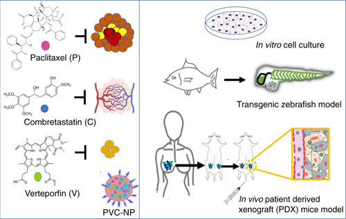
PDX: established TNBC PDX fragments were isolated from mice, and engrafted in mice again for in vivo transplantation and for in vitro organotypic slice culture. Biorender was used to construct part of the figure.

Acknowledgments
This image is the copyrighted work of the attributed author or publisher, and ZFIN has permission only to display this image to its users. Additional permissions should be obtained from the applicable author or publisher of the image. Full text @ Cell Death Dis.