
FIGURE SUMMARY
- Title
-
Neuromodulation: How Dopaminergic Neurons Shape and Modulate Behavior
- Authors
- Engert, F.
- Source
- Full text @ Curr. Biol.
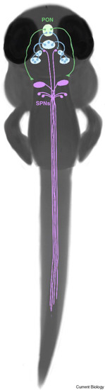
Figure 2. Dopamine neurons of larval zebrafish. Dopamine neurons in larval zebrafish form long-range projections from the preoptic nucleus (shown in green, close to the nose), and reciprocal connectivity between the posterior tuberculum (shown in blue centrally below the preoptic nucleus) and the intermediate and caudal nuclei of the hypothalamus (shown in blue at more lateral positions). The spinal projecting neurons of the reticulospinal array are depicted in purple. (Illustration: Anna Kehayova.) |
Acknowledgments
This image is the copyrighted work of the attributed author or publisher, and
ZFIN has permission only to display this image to its users.
Additional permissions should be obtained from the applicable author or publisher of the image.
Full text @ Curr. Biol.