
- Title
-
An Optical Illusion Pinpoints an Essential Circuit Node for Global Motion Processing
- Authors
- Wu, Y., Dal Maschio, M., Kubo, F., Baier, H.
- Source
- Full text @ Neuron
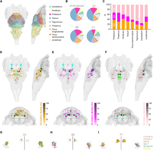
MAE-Correlated Neurons Are Clustered in the Ventral Lateral Pretectum (A) Previously annotated anatomical masks registered onto the standard brain. (B) Anatomical distribution of OMT subtypes in comparison with all DS neurons in the imaged volume. (C) Percentage of OMT subtypes in various brain areas. (D–F) Spatial distribution of OMT subtypes. Top left, top view; top right, side view from the right; bottom, frontal view. Relative density is calculated as the number of neighbors belonging to the same cluster in a radius of 20 μm, normalized by the highest density per subtype. Cyan dashed lines encircle the MAE cluster; green dashed lines encircle the non-MAE cluster. (G–I) Spatial distribution and tuning of OMT neurons within the MAE and the non-MAE cluster. Shown is a frontal view of the region marked by black dashed squares in (D)–(F). The dashed line represents the midline. Bi, binocular; Rot-sel, rotation-selective. See also Figures S5–S7. |