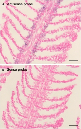
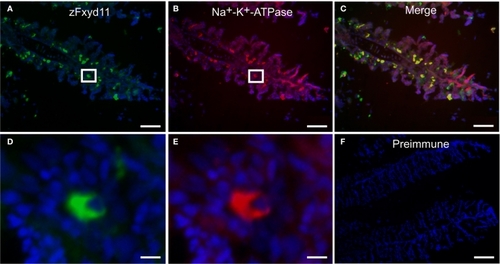
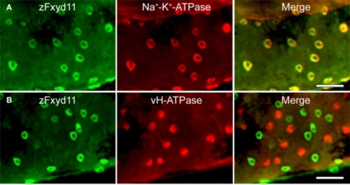
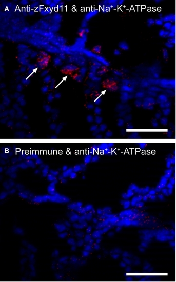
ZFIN is incorporating published figure images and captions as part of an ongoing project. Figures from some publications have not yet been curated, or are not available for display because of copyright restrictions.

EXPRESSION / LABELING:
Genes:
Fish:
Anatomical Terms:
Stage: Adult

ZFIN is incorporating published figure images and captions as part of an ongoing project. Figures from some publications have not yet been curated, or are not available for display because of copyright restrictions.

EXPRESSION / LABELING:
Gene:
Fish:
Conditions:
Anatomical Terms:
Stage Range: 5-9 somites to Adult
PHENOTYPE:
Fish:
Conditions:
Observed In:
Stage Range: Days 7-13 to Adult

Expression pattern of zfxyd11 mRNA in the gill of adult zebrafish. In situ hybridization was performed on paraffin-embedded gill sections of adult zebrafish with a DIG-labeled zfxyd11 antisense probe (A) or a sense probe as a control (B). Signals for zfxyd11 mRNA (violet) appeared to be abundant in the basal regions of the secondary lamellae. Scale bars, 10 μm.

EXPRESSION / LABELING:
Gene:
Fish:
Anatomical Term:
Stage: Adult

Localization of zFxyd11 in NaK-MRCs of the gill of adult zebrafish. Immunofluorescence staining was performed on cryosections of the gill of adult zebrafish with anti-Na+–K+-ATPase antiserum [red in (B), (C), (E)] along with either anti-zFxyd11 antiserum [green in (A), (C), (D)] or with preimmune serum [green in (F)]. Nuclei were stained with Hoechst 33342 (blue). High magnification images of zFxyd11-positive cells in the boxed areas in (A,B) are shown in (D,E), respectively. Scale bars in (A–C), 50 μm; in (D,E), 5 μm; in (F), 100 μm.

EXPRESSION / LABELING:
Gene:
Antibodies:
Fish:
Anatomical Terms:
Stage: Adult

Localization of zFxyd11 in NaK-MRCs of the skin of zebrafish larvae. Immunofluorescence staining was performed on 2-dpf zebrafish larvae with anti-zFxyd11 antiserum [green in (A), (B)], along with anti-Na+–K+-ATPase (red in (A)] or anti–vH+-ATPase antiserum [red in (B)]. Regions of the yolk sac extension are shown. Note that signals for zFxyd11 entirely overlap with NaK-MRCs (A), and are not colocalized with vH-MRCs (B). Scale bars, 40 μm.

EXPRESSION / LABELING:
Gene:
Antibodies:
Fish:
Anatomical Terms:
Stage Range: Long-pec to Adult

Interaction between zFxyd11 and Na+–K+-ATPase. In situ PLA was performed on cryosections of the gills of adult zebrafish with anti-Na+–K+-ATPase antiserum along with either anti-zFxyd11 antiserum (A) or corresponding preimmune serum (B). Nuclei were stained with Hoechst 33342 (blue). Signals of interaction of two proteins were detected on the basal regions of the secondary lamellae (red, arrows). Scale bars, 25 μm.

EXPRESSION / LABELING:
Gene:
Antibodies:
Fish:
Anatomical Terms:
Stage: Adult

Effects of zFxyd11 knockdown. (A–C) Immunofluorescence staining was performed on zfxyd11-MO-injected (A), control-MO-injected (B), and zfxyd11-MO with zfxyd11a mRNA-injected larvae (C) with anti-zFxyd11 antiserum (green), along with anti–Na+–K+-ATPase antiserum (red). No signal for zFxyd11 was detected in zfxyd11 morphants (A). (D) The number of Na+–K+-ATPase–positive cells in the skin was counted and is represented as the mean ± SEM. Significant differences were evaluated by one-way ANOVA, Tukey's multiple comparison test (***p < 0.001, n = 7). (E) Immunofluorescence staining was performed on zfxyd11-MO-injected (left panel), control-MO-injected larvae (right panel) with anti–vH+-ATPase antiserum. (F) The number of vH+-ATPase–positive cells in the skin was counted and is represented as the mean ± SEM. The number of vH+-ATPase-positive cells was not affected by zFxyd11 knockdown (n = 7).

EXPRESSION / LABELING:
Gene:
Antibodies:
Fish:
Knockdown Reagent:
Anatomical Terms:
Stage: Long-pec
PHENOTYPE:
Fish:
Knockdown Reagent:
Observed In:
Stage Range: High-pec to Long-pec

Membrane topology of zFxyd11a. (A) Homogenates of 293T cells expressing zFxyd11a-FLAG were fractionated into the cytosolic (lane 9) and membranous fractions. The membranes were incubated with PBS (lanes 1 and 2), 1 M NaCl (lanes 3 and 4), 0.1 M Na2CO3 (pH 11.5, lanes 5 and 6), or 1% Triton X-100 (lanes 7 and 8) followed by ultracentrifugation to separate soluble protein supernatants (S; lanes 1, 3, 5, and 7) from membranous pellets (P; lanes 2, 4, 6, and 8). The samples were analyzed by Western blotting with antibodies against FLAG, VTI1a (a TM protein), and DRP1 (a cytosolic protein). (B) HeLa cells were transfected with either zFxyd11a-FLAG (top panels) or FLAG-MHC-I (bottom panels) and were then processed for immunofluorescence staining with anti-FLAG antibody (red) without (left panels) or with (right panels) membrane permeabilization. Nuclei were stained with Hoechst 33342 (blue). Scale bars, 20 μm. (C) HeLa cells transfected with zFxyd11a-FLAG were lysed directly (Control) or were subjected to cell-surface biotinylation (Biotinylated). Whole cell lysates (left panels) and biotinylated samples (right panels) were analyzed by Western blotting with anti-FLAG antibody (top panels) and anti-DRP1 antibody (bottom panels).

Localization of zFxyd11 in NaK-MRCs of the skin of zebrafish larvae. Immunofluorescence staining was performed on 2-dpf zebrafish larvae with anti-zFxyd11 antiserum [green in (A–D)], along with anti–Na+–K+-ATPase [red in (A), (B)] or anti–vH+-ATPase antiserum [red in (C), (D)]. Control staining was carried out with preimmune serum (E,F). Regions of the yolk sac (A,C,E) and the yolk sac extension and the trunk (B,D,F) are shown. Note that signals for zFxyd11 entirely overlap with NaK-MRCs on the skin of yolk, yolk extension, and trunk (A,B), and are not colocalized with vH-MRCs (C,D). Scale bars, 200 μm.

Acknowledgments
This image is the copyrighted work of the attributed author or publisher, and ZFIN has permission only to display this image to its users. Additional permissions should be obtained from the applicable author or publisher of the image. Full text @ Front. Physiol.