FIGURE SUMMARY
Title

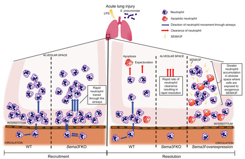
Semaphorin 3F signaling actively retains neutrophils at sites of inflammation

Authors
Plant, T., Eamsamarng, S., Sanchez-Garcia, M.A., Reyes, L., Renshaw, S.A., Coelho, P., Mirchandani, A.S., Morgan, J.M., Ellett, F.E., Morrison, T., Humphries, D., Watts, E.R., Murphy, F., Raffo-Iraolagoitia, X.L., Zhang, A., Cash, J.L., Loynes, C., Elks, P.M., Van Eeden, F., Carlin, L.M., Furley, A.J.W., Whyte, M.K.B., Walmsley, S.R.
Source
Full text @ Journal of Clin. Invest.

Inflammatory human neutrophils express SEMA3F and its coreceptor NRP2.

(A and B) Lung sections taken at time of tumor resection from nontumor regions of patients with moderate severity COPD were stained for SEMA3F or NRP 2 (A), or a combination of CD66b (green), NRP2 (red), and DAPI (blue) (B). Images taken at ×40 magnification. Scale bars: 100 μm in A, 20 μm in B. Human blood neutrophil SEMA3F protein expression following 4 hours culture ex vivo was assessed by Western blot (C), and fold change to unstimulated control was determined by densitometry normalized to P38 (D). The percentage of blood monocytes (CD66b, CD14/49D+) and neutrophils (CD66b+) expressing NRP1 and NRP2 was determined in freshly isolated cells (E) and following ex vivo culture for 4 hours by flow cytometry in control and stimulated conditions (F). Data are mean ± SEM, with individual data points (n = 3–5) from independent experiments. Human blood neutrophil NRP2 protein expression following 4 hours culture ex vivo was assessed by Western blot (G) and fold change to unstimulated control was determined by densitometry normalized to P38 (H). Statistical analysis: 1-way ANOVA and Bonferroni’s post hoc tests (D and H) and 2-way ANOVA and Sidak’s post hoc tests (E and F) were performed. *P < 0.05; **P < 0.01; ***P < 0.001.

Knockdown of semaphorin 3F by MO injection or by TALEN-induced mutation accelerates resolution of neutrophilic inflammation in zebrafish.

(AD) sema3fa and/or sema3fb MO (1 nL of 0.5 mM) was injected into 1-cell-stage zebrafish mpx:GFP embryos, with 1 nL of 0.5 mM control MO used as a negative control. Tail fin transection was performed at 2 dpf, and neutrophils were counted at 6 hpi and 24 hpi. (A) Neutrophil counts at the 6 hpi time point with (B) overlaid fluorescence and bright-field photomicrographs (recruitment). (C) Neutrophil counts at the 24 hpi time point with (D) overlaid fluorescence and bright-field photomicrographs (resolution). Scale bars: 60 μm (B and D). Data are mean ± SEM, with individual data points (n = 30) from 3 independent experiments. (EG) sema3fa- or sema3fb-mutated F1 fish were incrossed and compared with mpx:GFP fish. Tail fin transection was performed at 2 dpf, and (E) neutrophils were counted at 6 hpi. Data are mean ± SEM, with individual data points (n = 30) from 4 independent experiments. (F) Whole-body total neutrophil numbers were counted at 3 dpf. Data are mean ± SEM, with individual data points (n = 60) from 4 independent experiments. (G) Tail fin transection was performed at 2 dpf and neutrophils were counted at 24 hpi. Data are mean ± SEM, with individual data points (n = 5–60) from 4 independent experiments. (H) sema3fa and or sema3fb MOs (1 nL of 0.5 mM) were injected into 1-cell-stage zebrafish mpx:kaede embryos, and tail fin transection was performed at 2 dpf. Neutrophils at 6 hpi were recruited to the wound and photoconverted, and red fluorescence neutrophils were tracked for 3.5 hours. Data are from 3 independent experiments (n = 9). Statistical analysis was by 1-way ANOVA and Bonferroni’s post hoc test. ***P < 0.001.

Neutrophil-specific loss of <italic>Sema3f</italic> results in more rapid neutrophil recruitment to and clearance from the lungs in a murine acute lung injury model.

(A and B) Fold change in Sema3f and Nrp2 gene expression following acute lung injury with LPS. Mice were sacrificed at 6, 24, and 48 hours after instillation. BAL neutrophils were collected and cDNA was extracted. TaqMan analysis of cDNA was performed with data normalized to murine Actb gene expression. Data are mean ± SEM of fold change compared with peripheral blood neutrophils (T0 PB) from 2 individual experiments (n = 4–6). An acute lung injury was induced by intratracheal LPS instillation, mice were sacrificed at 24 hours, and lung sections were stained for expression of the Ly6G neutrophil marker and SEMA3F (C), NRP1, and NRP2 (D). Scale bars: 50 μm. (E and F) Sema3ffl/flMrp8Cre–/– (WT) and Sema3ffl/flMrp8Cre+/– (KO) mice were challenged with LPS, sacrificed at 2, 6, 24, and 48 hours, and BAL fluid was obtained. Cell counts were performed by hemocytometer and the differential cell count was established by cytospins. Time to 50% reduction in peak neutrophil number was calculated individually for each genotype (T50) (E). BAL fluid IgM content was measured by ELISA. Data are shown as log-transformed fold change from WT (F). Apoptosis was assessed by morphology, with data as mean ± SEM (G) from 3 individual experiments (n = 6–12). Statistical analysis was by 1-way ANOVA and Bonferroni’s post hoc test (A and B) and 2-way ANOVA with Sidak’s post hoc test (EG). *P < 0.05; **P < 0.01; ***P < 0.001.

Neutrophil-driven deletion of <italic>Sema3f</italic> favors a selective allocation of neutrophils in the alveolar space while retaining antimicrobial capacity.

(A and B) Sema3ffl/flMrp8Cre–/– (WT) and Sema3ffl/flMrp8Cre+/– (KO) mice were challenged with nebulized LPS and sacrificed at 0, 2, and 6 hours following LPS challenge. Blood and lung tissues were harvested with lung digest for Ly6G staining (neutrophil number). In a parallel series of experiments, lungs were instilled with agarose gel at 6 hours, then fixed and stained with the endothelial marker CD31 (green) and the neutrophil marker S100A9 (red). Lungs were imaged by confocal microscopy (Zeiss LSM 880 with Airyscan) with 3D reconstruction and neutrophil position relative to the blood vessels assigned, using Imaris software version 9.1 (neutrophil, white arrows) (C). Percentage of total neutrophils per 106 μm3 lung tissue is shown, with a minimum of 190 neutrophils quantified per mouse (D). Data are mean ± SEM from 3 individual experiments (n = 3–5). (E and F) WT and KO mice were challenged with intratracheal instillation of S. pneumoniae, and lung bacterial counts (E) and BAL and lung neutrophil counts (F) were undertaken 14 hours after challenge. Data are mean ± SEM from 2 individual experiments (n = 6–10). Statistical analysis was by 2-way ANOVA with Sidak’s post hoc test (A, B, and D) and Mann-Whitney (E) and 1-tailed unpaired t test (F). *P < 0.05.

Overexpression of <italic>sema3f</italic> in a zebrafish model of inflammation delays neutrophil recruitment and resolution of the inflammatory response.

(AD) sema3fa or sema3fb RNA (50 ng/μL) was injected into 1-cell-stage zebrafish mpx:GFP embryos, with 50 ng/μL mCherry RNA used as a negative control. Tail fin transection was performed at 2 dpf, and neutrophils counted at 6 and 24 hpi. (A) Neutrophil counts at the 6 hpi time point with (B) overlaid fluorescence and bright-field photomicrographs (recruitment). (C) Neutrophil counts at the 24 hpi time point with (D) overlaid fluorescence and bright-field photomicrographs (resolution). Scale bars: 60 μm (B and D). Data are mean ± SEM with individual data points from 3 independent experiments (n = 30). (E) sema3fa and/or sema3fb RNA (50 ng/μL) were injected into 1-cell-stage zebrafish mpx:kaede embryos, and tail fin transection was performed at 2 dpf. Neutrophils recruited to the wound at 6 hpi were photoconverted, and red fluorescent neutrophils were tracked for 3.5 hours. Data are mean ± SEM from 3 independent experiments (n = 9). (F and G) sema3fa or sema3fb RNA (50 ng/μL) was injected into 1-cell-stage zebrafish mpx:GFP embryos, with 50 ng/μL mCherry RNA used for control. Tail fin transection was performed at 2 dpf. Neutrophil movement was tracked over 1 hour by time-lapse microscopy during the recruitment phase of inflammation (1–2 hpi) and speed of neutrophil migration (F) and meandering index (displacement/path length) (G) were determined. Data are mean ± SEM from 3 independent experiments (each point represents a single neutrophil) (n = 15). (H) sema3fa or sema3fb RNA (50 ng/μL) was injected into 1-cell-stage Tg(lyz:PHAkt-EGFP) embryos with noninjected controls, tail fin transection was performed at 2 dpf, and polarity indices were calculated for neutrophils recruited to the tail region. Data are mean ± SEM from a single experiment (n = 8). Statistical analysis was by 1-way ANOVA and Bonferroni’s post hoc test. ***P < 0.001.

Exogenous SEMA3F retains recruited neutrophils at the injury site in a murine model of acute lung injury.

(A and B) Intratracheal (IT) recombinant SEMA3F (1 μM) was administered to C57BL/6 mice 24 hours after nebulized LPS challenge or PBS. Mice were then sacrificed at 48 and 72 hours and BAL was performed with differential apoptosis cell/neutrophil counts (A), or lungs were retained for fixed lung slice imaging (B). Lungs harvested for lung imaging were instilled with agarose gel, and fixed and stained with the endothelial marker CD31 (green) and the neutrophil marker S100A9 (red). Lungs were imaged by confocal microscopy (Zeiss LSM 880 with Airyscan) with 3D reconstruction and neutrophil position relative to the blood vessels was assigned using Imaris software version 9.1, with at least 80 neutrophils quantified per mouse. Data are mean ± SEM with individual data points from 4 independent experiments (n = 12). (CE) Naive Catchup (IVM-RED;Lifeact-GFP) mice were sacrificed and lungs were instilled with agarose gel, precision sliced, and imaged by confocal microscopy for 90 minutes with addition of SEMA3F or PBS vehicle control at 30 minutes. Following treatment, neutrophil mean speed (C), maximum speed (D), and track straightness (directionality) (E) were measured and analyzed for 60 minutes using Imaris software version 9.1. Data are from 3 independent experiments (n = 3). Statistical analysis was by 2-way ANOVA and Sidak’s post hoc test (A and B) or paired t test (CE). *P < 0.05; **P < 0.01; ****P < 0.0001.

Neutrophil treatment with exogenous SEMA3F blocks chemotactic responses while preserving phagocytic capacity and respiratory burst functions.

(AF) Isolated peripheral blood neutrophils from healthy volunteers were incubated with recombinant SEMA3F (0–100 nM), and functional assays were performed. (A and B) Chemotactic behavior of neutrophils to fMLF (0–100 nM) and LTB4 were measured by Boyden chamber, CC chemokinesis control (A), and microfluidic chip assay (B). (C) Neutrophils were incubated with Alex 488 E. coli and phagocytic uptake was determined by flow cytometry, with adhesion excluded by 4°C control. (D) Phagocytic indices were calculated by cytospin following neutrophil culture with opsonized Zymosan particles for 30 minutes. (E) ROS generation was determined following a 1 hour preincubation with SEMA3F and treatment with fMLF for 30 minutes. (F) Neutrophil release of elastase was measured by fluorimetric assay following pretreatment with 1 hour of SEMA3F, 30 minutes of GM-CSF (10 ng/mL), and 10 minutes of fMLF (100 nM). All data are mean ± SEM with individual data points from independent experiments (n = 3–6). Statistical analysis was by 1-way ANOVA and Sidak’s post hoc test (A, B, E, and F) or paired t test (CE). *P < 0.05; **P < 0.01; ****P < 0.0001.

SEMA3F promotes neutrophil rounding and F-actin disassembly.

(AC) sema3fa or sema3fb RNA (50 ng/μL) was injected into 1-cell-stage Tg(mpx:LifeActRuby) embryos, tail fin transection was performed at 2 dpf, and neutrophil fluorescence intensities were calculated (A). Representative images (B). White arrows indicate the direction to the wound edge. Roundness scores were calculated for neutrophils recruited to the injury (C). Data are mean ± SEM from an single experiment (n = 9). (DG) Human blood neutrophils were pretreated with PBS or 100 nM SEMA3F before stimulating with PBS or 100 nM fMLF. Following DAPI/cell mask/phalloidin staining, neutrophil rounding was quantified by high-content widefield microscopy (1 = perfect sphere), with more than 1000 cells measured per condition (D). Pixel intensity for phalloidin was obtained using confocal microscopy (×100 objective), scale bars: 5 μm (E). F-actin cell content was quantified with more than 1000 cells measured per condition (F and G) and distribution was used to calculate a polarity index (H). Data are mean ± SEM from 4 independent experiments (n = 4–22). (IK) Intratracheal SEMA3F (1 μM) was administered to C57BL/6 mice 24 hours after nebulized LPS or PBS, and lung tissue (I and J) or BAL fluid (K) was collected at 48 hours. Lungs were instilled with agarose gel, fixed and stained with the endothelial CD31 and the neutrophil marker S100A9, and imaged by confocal microscopy. The percentage of neutrophils of each sphericity (I) and the mean sphericity of neutrophils (J) was defined. F/G actin ratios per cell were calculated from fluorescence intensities following staining with phalloidin (F-actin) and DNAse 1 (G-actin), with a latrunculin B (LTNB) negative control (K). Data are mean ± SEM from 2 independent experiments (n = 9) (IK). Statistical analysis was by 1-way ANOVA and Bonferroni’s post hoc test (AD and H), with Sidak-Holm multiple comparison posttest (K) and paired t test (G) performed for time points 15 to 30 minutes during the steady state of F-actin turnover, and unpaired t test (IJ). *P < 0.05, **P < 0.01, ***P < 0.001, ****P < 0.0001.

A summary of the migratory movement of neutrophils in <italic>Sema3f</italic> knockdown and overexpression models of acute lung injury.

Neutrophil-specific KO of Sema3f increased neutrophil transit through the airways and led to more rapid clearance of neutrophils from the alveolar space, resulting in faster resolution. In contrast, overexpression of Sema3f in the airspaces caused neutrophil retention and delayed inflammation resolution.

Acknowledgments
This image is the copyrighted work of the attributed author or publisher, and ZFIN has permission only to display this image to its users. Additional permissions should be obtained from the applicable author or publisher of the image. Full text @ Journal of Clin. Invest.