FIGURE SUMMARY
Title

Loss of Crb2b-lf leads to anterior segment defects in old zebrafish

Authors
Kujawski, S., Crespo, C., Luz, M., Yuan, M., Winkler, S., Knust, E.
Source
Full text @ Biol. Open

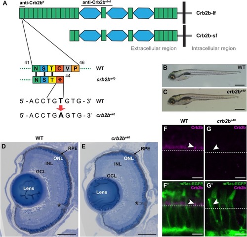
crb2be40 mutants are homozygous viable and develop normal eyes. (A) Schematic illustration of the long and short isoforms of Crb2b, Crb2b-lf and Crb2b-sf. Green rectangles, EGF-like domains; light blue hexagons, domains with similarity to the globular domain of laminin A (LamG domain). Bars above the protein indicate the regions used as antigens for anti-Crb antibodies. In the crb2be40 allele, a T to A transversion translates to an early stop codon at amino acid position 44 of the protein, resulting in truncated Crb2b-lf. (B,C) Brightfield images of WT (B) and crb2be40 (C) zebrafish larvae at 5 dpf. Mutant larvae have overall normal appearance. (D,E) Transverse retinal sections stained with Toluidine Blue show normal lamination of the retina of crb2be40 (E) larvae in comparison to WT (D) at 5 dpf. Asterisk denotes the optic nerve. GCL, ganglion cell layer; INL, inner nuclear layer; ONL, outer nuclear layer; RPE, retinal pigment epithelium. (F–G′) Confocal images of transverse retinal sections of larvae at 3 dpf stained with rabbit anti-Crb2be8e9 in the Tg(bactin:mRas-EGFP) background. Crb2b is detected in the IS of WT PRCs (F,F′, white arrowheads), but not in crb2be40 mutant PRCs (G,G′). White dashed lines mark the position of the outer limiting membrane. Scale bars: (B,C) 1 mm; (D,E) 100 µm; (F–G′) 5 µm.

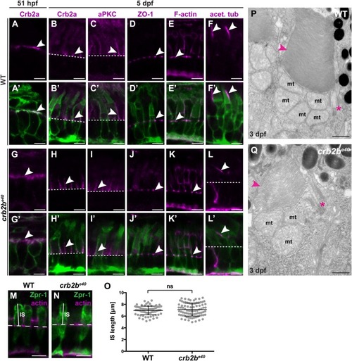
crb2be40 mutant PRCs have normal morphology and polarity. (A–L′) Immunostaining of transverse sections of WT (A–F′) and crb2be40 (G–L′) retinas in the Tg(bactin:mRas-EGFP) background at 51 hpf (A,A′,G,G′) and 5 dpf (B–F′,H–L′). Crb2a localises on the entire apical membrane in WT (A,A′) and crb2be40 mutants (G,G′) PRC precursors of this stage (arrowheads). At 5 dpf, Crb2a and aPKC localise to the IS (arrowheads) in WT (B,B′,C,C′) and in mutant cells (H,H′,I,I′). ZO-1 and F-actin localise to the outer limiting membrane (OLM; white arrowheads), both in WT (D–E′) and mutants (J–K′). A well-formed axoneme (white arrowheads) is detected in retinal sections in both WT (F,F′) and crb2be40 (L,L′) fish by acetylated tubulin antibody staining. White dashed lines indicate OLM. (M,N) Transverse retinal sections of WT (M) and crb2be40 (N) larvae at 3 dpf stained with Zpr-1 antibody to mark the cell body of double-cone PRCs (green) and phalloidin to mark the OLM (magenta). The solid white lines indicate IS height, the dashed white lines mark the position of the OLM. (O) Quantification of IS length (µm) in both WT and crb2be40 PRCs. IS length was measured from the level of the OLM to the base of the OS (see M,N). At least 20 cells from three independent retinas were measured. Statistical significance was calculated by t-test (unpaired, with equal s.d., two-tailed). ns, not significant (P=0.3053). (P,Q) Transmission electron micrographs of WT (P) and crb2be40 (Q) OSs at 3 dpf. The overall ultrastructure of cilia (magenta asterisks) and OS membrane discs (magenta arrowheads) is preserved in mutant PRCs. mt, mitochondrium. Scale bars: (A–N) 5 µm; (P,Q) 1 µm.

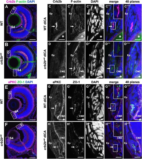
Crb2b is expressed in polarised cells of the ICA at 5 dpf. Immunostaining of transverse cryosections through the eye. (A,B,E,F) Overview of a WT (A,E) and a mutant (B,F) eye at 5 dpf. Higher magnifications of the boxed regions in the dorsal ICA are shown in C–D‴′ (WT) and G–H‴′ (crb2be40). Higher magnifications of the boxed regions in the ventral ICA are shown in Fig. S4. Crb2b staining (anti-Crb2be8e9) is detected in a cluster of cells in the WT ICA (C) but not in the mutant ICA (D). F-actin (C′,D′; visualised by phalloidin), aPKC (G,H) and ZO-1 (G′,H′) appear apically enriched (C′,D′). Arrows point to the cluster of polarised Crb2b expressing cells, and asterisks mark the lens. dICA, dorsal iridocorneal angle; Ep, epidermis; CMZ, ciliary marginal zone. Scale bars: (A,B,E,F) 50 µm; (C–D‴′, G–H‴′) 10 µm.

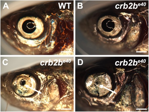
Old crb2be40 mutants have abnormal AS tissues. Brightfield images of old adult WT (A) and crb2be40 (B–D) eyes. Arrows in C and D point to a reduced pupil. Scale bars: 1 mm.

PHENOTYPE:
Fish:
Observed In:
Stage: Adult

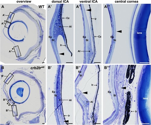
Loss of Crb2b-lf leads to an overgrowth of the iris in old fish. Toluidine Blue-stained transverse retinal sections of WT (A–A″′) and crb2be40 (B–B″′) adult zebrafish. (A–A″′) Overview of the whole WT eye (A) shows the normal appearance of the pupil (bordered by two asterisks). Higher magnification of WT dorsal (A′) and ventral (A″) iridocorneal angles shows the morphology of the iris, annular ligament cells and endothelial cells lining the ligament (arrowhead). A″′ shows the structure of the cornea, with the corneal endothelium cells being barely visible (A″′, arrowhead). (B–B″′) Overview of the whole eye of a mutant fish (B) demonstrates a decrease in pupil size (bordered by two asterisks) due to overgrowth of ICA tissue and an expansion of the iris towards the centre of the cornea (B″′). Multiple layers of cells line the posterior surface of the cornea (B″′, arrowhead). These cells appear to extend from the annular ligament (B′,B″ arrowheads). Ep, corneal epidermis; St, corneal stroma; Al, annular ligament; Ir, iris; Cz, ciliary zone. Scale bars: (A,B) 100 µm; (A′–A″′) and (B′–B″′) 50 µm.

PHENOTYPE:
Fish:
Observed In:
Stage: Adult

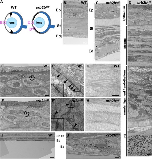
Loss of Crb2b-lf leads to corneal abnormalities in old fish. (A) Simplified schematic of a WT (left) and a crb2be40 (right) adult eye phenotype. Boxes denote regions of TEM images shown in B–D. (B–D) TEM transverse sections through the central cornea of adult WT (B) and crb2be40 fish (C,D) without (C) and with (D) iris expansion. (E–J) Higher magnification of the corneal layers in WT and mutant fish. Mutant corneal epidermal cells (F) do not appear different from their WT counterparts (E), and junctions (boxed regions in E,F) are visible in both (E′,F′, arrowheads). In the corneal stroma, collagen fibres are orthogonally layered both in the WT (G) and in the mutant (H). The endothelium is monolayered in WT (B,I), but appears as a multi-layered tissue in the mutant (C,J; labelled as annular ligament/endothelium in C), next to the elongated iris (D). Ep, corneal epidermis; St, corneal stroma; Ed, corneal endothelium. Scale bars: (B–F,I,J) 2 µm; (G,H) 0.5 µm.

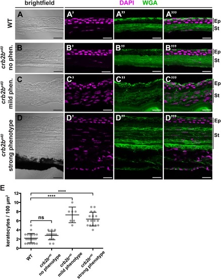
Loss of Crb2b-lf leads to an increase in the number of stromal keratocytes in old fish. (A–D‴). Brightfield (A,B,C,D), DAPI (magenta) and WGA (green) staining (A′–A″′,B′–B″′,C′-C″′,D′–D″′) of transverse cryosections of WT (A–A″′) and crb2be40 mutant (B–D″′) adult cornea. The stroma of crb2be40 mutants, which show an aberrant phenotype (C–D″′), appears to have more keratocyte nuclei (magenta) than mutants with no phenotype (B–B″′) or WT (A–A″′). Keratocyte nuclei were identified based on their flat and elongated morphology. Ep, corneal epidermis; St, corneal stroma. Scale bars: 5 µm. (E) Quantification of keratocyte nuclei in stromal tissue in WT versus crb2be40 mutant fish. The phenotypic categories correspond to the ones in A–D″′. Nuclei were counted from two separate 100 µm2 areas per adult fish in the central cornea, except for the ‘mild phenotype’ category due to lack of iris-free area at the centre of the cornea. Statistical significance was calculated by t-test (unpaired, with equal s.d., two-tailed). ****P<0.001; ns, not significant (P=0.0779).

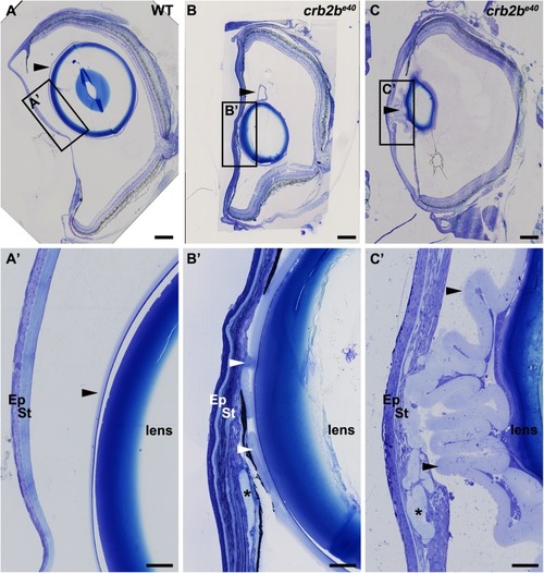
Loss of Crb2b-lf leads to lens capsule abnormalities in old fish. Toluidine Blue staining of transverse sections of WT (A,A′) and two crb2be40 mutant (B–C′) adult zebrafish eyes. In WT, the lens capsule is thin and smooth (A,A′, arrowheads) and is close to the lens. In mutant eyes (B–C′) the lens capsule appears thickened and folded (arrowheads). Asterisks mark areas where the lens capsule has penetrated into the cornea (B′,C′). Scale bars: (A–C) 200 µm; (A′–C′) 50 µm.

PHENOTYPE:
Fish:
Observed In:
Stage: Adult
Acknowledgments
This image is the copyrighted work of the attributed author or publisher, and ZFIN has permission only to display this image to its users. Additional permissions should be obtained from the applicable author or publisher of the image. Full text @ Biol. Open