
- Title
-
Galanin Signaling in the Brain Regulates Color Pattern Formation in Zebrafish
- Authors
- Eskova, A., Frohnhöfer, H.G., Nüsslein-Volhard, C., Irion, U.
- Source
- Full text @ Curr. Biol.

ZFIN is incorporating published figure images and captions as part of an ongoing project. Figures from some publications have not yet been curated, or are not available for display because of copyright restrictions. |
|
ZFIN is incorporating published figure images and captions as part of an ongoing project. Figures from some publications have not yet been curated, or are not available for display because of copyright restrictions. |
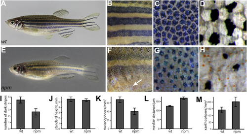
The (A–D) Wild-type adult zebrafish (A), magnified mid-trunk area (B), expanded melanophores (C), and detail of 1st ventral dark stripe in fixed specimen (D). (E–H) (I and J) Number of dark stripes (I) in fish of comparable height (J) (mean ± SD, n = 10). (K) Density of melanophores (mean ± SD, n = 10). (L) Distance between melanophores in the 1st ventral stripe (median ± SD, n = 10). (M) Density of xanthophores in the first light stripe (mean ± SD, n = 10). PHENOTYPE:
|
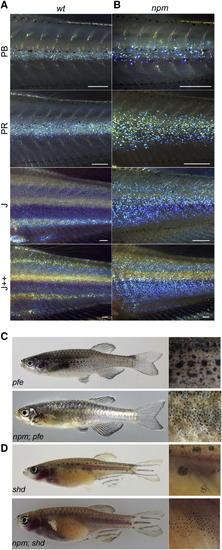
Pigment Pattern of (A) Wild-type iridophores appear as a dense sheet along the horizontal myoseptum (stages PB and PR); they spread dorsally and ventrally where they form secondary light stripes (stages J and J++) [ (B) In (C) (D) PHENOTYPE:
|
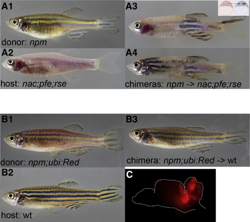
(A) Chimeric animals (A3, A4) derived from blastomere transplantations of (B) Blastomere transplantations of (C) Fluorescent image of an open brain (outlined). |
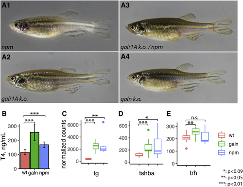
(A) Phenotypes of (B) T4 thyroid hormone levels measured in wild-type, (C–E) Relative transcript abundance for |