
- Title
-
Involvement of an Oct4-related PouV gene, pou5f3/pou2, in neurogenesis in the early neural plate of zebrafish embryos
- Authors
- Inomata, C., Yuikawa, T., Nakayama-Sadakiyo, Y., Kobayashi, K., Ikeda, M., Chiba, M., Konishi, C., Ishioka, A., Tsuda, S., Yamasu, K.
- Source
- Full text @ Dev. Biol.
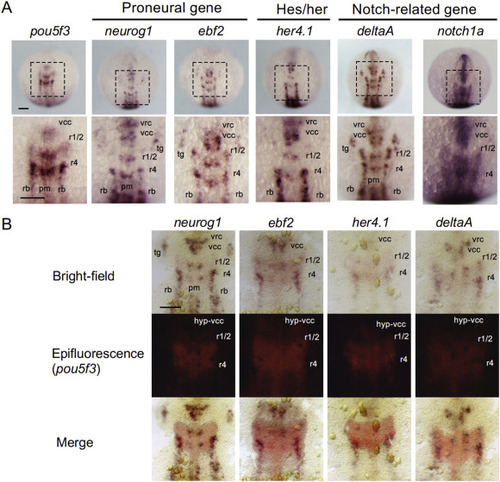
Comparison of the expression patterns of pou5f3 and neurogenesis genes in the neural plate. A. The expression of pou5f3 and various neurogenesis genes, including proneural genes (neurog1, ebf2), a her gene (her4.1), and Notch-related genes (deltaA, notch1a), in the neural plate was compared at the bud stage using WISH. Dorsal views with anterior to the top. Top row, dorsal views of whole embryos; bottom row, enlarged views of the areas marked with broken lines in the top row. B. The expression of pou5f3 (red) and neurogenesis genes (brown) in the midbrain-hindbrain region of the neural plate was compared by two-color in situ hybridization at the bud stage. Top row, bright-field views; middle row, epifluorescence views; bottom row, merged views. pm, primary motor neuron progenitor; rb, primary sensory neuron progenitors (Rohon-Beard neurons); r1/2, medial and lateral neuron progenitors in r1/2; r4, medial and lateral neuron progenitors in r4; tg, trigeminal ganglion; vcc, ventrocaudal cluster; vrc, ventrorostral cluster. Scale bar, 100 μm. EXPRESSION / LABELING:
|
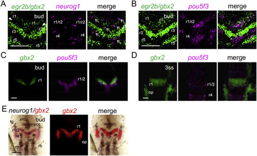
Close association of the expression domains of neurog1 and pou5f3 in rhombomeres 1, 2, and 3. A–D. The relationships between the expression domains of neurog1 and pou5f3 in the neural plate were compared by two-color FISH with those of gbx2 and egr2b in r1 and r3/r5, respectively, at the bud (A–C) and three-somite (D) stages. Expression gaps between the gbx2 domain in r1 and egr2b domain in r3 are marked with arrowheads, and neurog1 domains are marked with arrows (A). (E) The expression of neurog1 and gbx2 was compared by conventional two-color WISH. Dorsal views with anterior to the top. For abbreviations, see the legend for Fig. 1. Scale bar, 100 μm. EXPRESSION / LABELING:
|
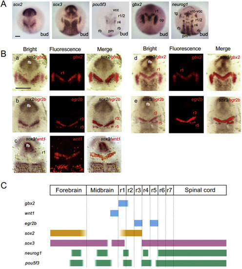
Localization of the proneural clusters and soxB1 regions in the neural plate. A. Expression of soxB1, pou5f3, gbx2, and neurog1 at the bud stage revealed by single-color WISH. B. Comparison of the gene expression patterns by two-color WISH. The expression domains of two specified genes are stained brown and red. The bright-field views (left), epifluorescence views (middle), and merged views (right) are shown for each of the gene pairs examined. fb, forebrain. For the other abbreviations, see the legend for Fig. 1. Scale bars, 100 μm. C. Schematic views of the expression domains of brain regional marker genes, soxB1, and neurog1/pou5f3. EXPRESSION / LABELING:
|
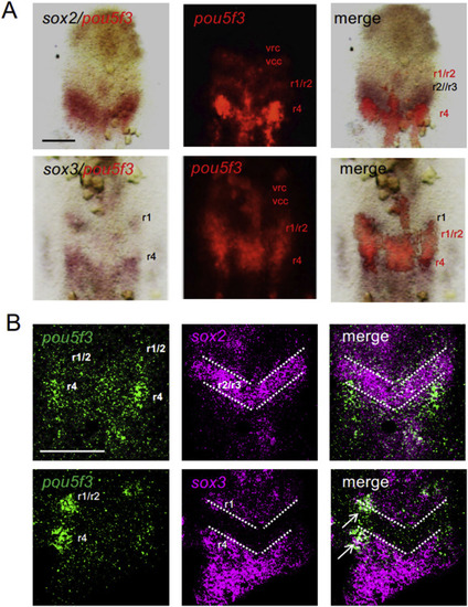
Relationship between the expression regions of pou5f3 and soxB1. A. Expression of soxB1 (brown) and pou5f3 (red) was compared at the bud stage by two-color WISH. Bright-field views (left), epifluorescence views (middle), and merged views (right) are shown for each of the gene pairs examined. B. Expression of soxB1 (magenta) and pou5f3 (green) was compared at the bud stage by two-color FISH. White dotted lines show rhombomere boundaries. The right side of the hindbrain is out of focus in the bottom row due to tilted mounting. For abbreviations, see the legend for Fig. 1. Scale bars, 100 μm. EXPRESSION / LABELING:
|

ZFIN is incorporating published figure images and captions as part of an ongoing project. Figures from some publications have not yet been curated, or are not available for display because of copyright restrictions. EXPRESSION / LABELING:
PHENOTYPE:
|
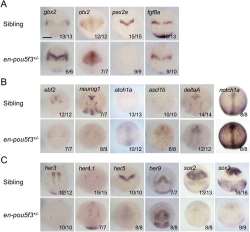
Effects of functional suppression of pou5f3 on the expression patterns of brain-forming genes. Embryos from crosses of wild-type and en-pou5f3+/− Tg fish were exposed to heat shock at 90% epiboly for 1 h at 37 °C. They were then examined to elucidate the expression of brain regionalization genes (A) and neurogenesis genes (B, C) by WISH and genotyped. Dorsal views with anterior to the top are shown. The numbers of embryos exhibiting the patterns shown and those of stained embryos are shown at the right bottom. Scale bars, 200 μm. |
Reprinted from Developmental Biology, 457(1), Inomata, C., Yuikawa, T., Nakayama-Sadakiyo, Y., Kobayashi, K., Ikeda, M., Chiba, M., Konishi, C., Ishioka, A., Tsuda, S., Yamasu, K., Involvement of an Oct4-related PouV gene, pou5f3/pou2, in neurogenesis in the early neural plate of zebrafish embryos, 30-42, Copyright (2019) with permission from Elsevier. Full text @ Dev. Biol.