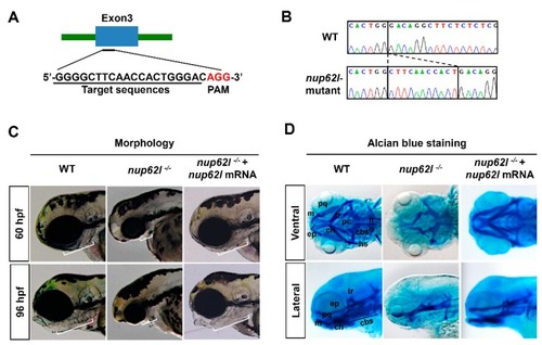
Loss of Nup62l led to severely impaired formation of PA cartilages. (A) Diagram showing the CRISRP/Cas9 target DNA sequence (underline) of zebrafish gene nup62l. PAM region (AGG) was shown in red. (B) Sanger sequencing results revealed an 11-bp genomic DNA fragment insertion at the target site in nup62l-mutants. (C) Lateral views showing the pharyngeal morphology of WT siblings. Embryos of nup62l-mutants at 60 hpf or 96 hpf were injected with or without 300 pg nup62l mRNA/embryo. White lines indicated the pharyngeal regions. Note a shortened and thinned pharyngeal region in nup62l-mutants. (D) Images showing pharyngeal cartilage elements stained with alcian blue in WT. The nup62l-mutants injected with or without 300 pg nup62l mRNA at 96 hpf were ventrally viewed in upper panels and laterally viewed in lower panels. cbs, ceratobranchials; ch, ceratohyal; ep, ethmoid plate; hs, hyosymplectic; m, Meckel’s cartilage; no, notochord; pc, parachordal; pq, palatoquadrate; tr, trabecula.

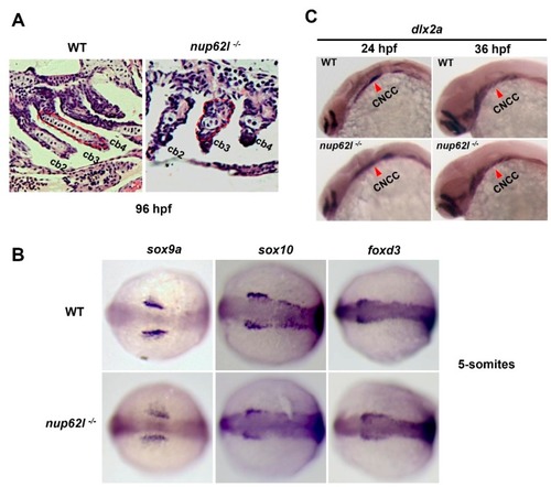
Specification and migration of CNCCs were unperturbed by loss of Nup62l. (A) Representative images showing the severely impaired PA chondrocytes of nup62l-mutants on sections stained with hematoxylin and eosin at 96 hpf. The 2nd–4th ceratobranchials (cb) were indicated. (B) Dorsal views with anterior region toward the left of 5-somites embryos stained with early crest markers (sox9a, sox10 and foxd3). (C) WT and nup62l-mutant embryos were stained with probes of dlx2a, a marker of CNCCs migration at 24 hpf and 36 hpf. Lateral views of CNCCs as pointed by red arrowheads.

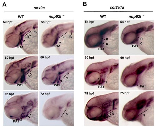
Nup62l was essential for condensation and differentiation of pharyngeal chondrogenic progenitors. WISH expression patterns of markers sox9a (A) and col2a1a (B) in nup62l-mutants at indicated stages. Embryos in (A) and (B) were shown as lateral views with anterior to the left. Question markers indicate the pharyngeal regions with no or unrecognized PA. PA, pharyngeal arches; fb, fin bud; nc, neurocranium.

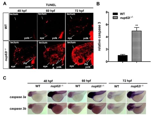
Loss of Nup62l induced extensive apoptosis in the impaired PA region. (A) Detection of apoptotic cells in PA of nup62l-mutants with TUNEL assays at indicated stages. PA, pharyngeal arches. (B) Strongly elevated caspase 3 activity in nup62l-mutants at 60 hpf in comparison with that in WT siblings. The data expressed as Mean ± SD were representatives of three independent experiments, each done with three samples of 15 zebrafish. ** p < 0.01. (C) WISH assays showing lateral views of the expression of caspase 3a and caspase 3b genes in WT or nup62l-mutants at 48 hpf, 60 hpf or 72 hpf.

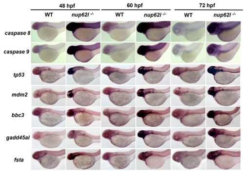
Loss of Nup62l activated intrinsic and extrinsic apoptotic pathways. WISH assays showing lateral views of the expression of various apoptosis-related genes (caspase 8, caspase 9, tp53/p53, mdm2, bbc3, gadd45al and fsta) in WT siblings and nup62l-mutants at different stages.

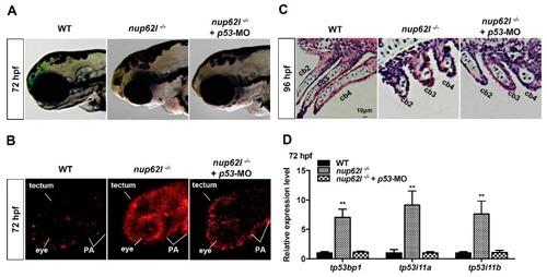
Activation of p53-dependent apoptotic pathway contributed to the defective formation of PA in nup62l-mutants. (A) Partial rescue of morphological defects in PA of nup62l-mutants injected with 5 ng p53-MO as shown by the lengths of white lines. (B) TUNEL assays were performed to assess the apoptosis in pharyngeal region of nup62l- and p53- double inactivated mutants compared to WT and nup62l-mutants at 72 hpf. Embryos were imaged by confocal microscopy. PA region, eyes and optic tectum were labeled. (C) PA sagittal sections at 96-hpf of WT, nup62l-mutants or nup62l-mutants injected with 5 ng p53-MO were analyzed with hematoxylin and eosin staining. The 2nd-4th ceratobranchials (cb) were labeled. (D) Expression levels of genes (tp53bp1, tp53i11a and tp53i11b) implicated in p53-dependent apoptotic pathway were analyzed by qRT–PCR in WT, nup62l-mutants or nup62l-mutants injected with 5 ng p53-MO at 72 hpf. Expression levels were normalized to WT embryos. Data expressed as Mean ± SD were representatives of three independent experiments. ** p < 0.01.

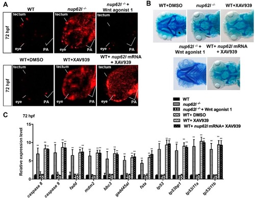
Suppression of Wnt/β-catenin signaling by Nup62l deprivation activated multiple apoptotic pathways. (A) Apoptotic cells were detected by TUNEL assays at 72 hpf in PA of WT embryos, nup62l-mutants, nup62l-mutants treated with 10 μM Wnt agonist 1, WT treated with 0.15% DMSO or 15 μM XAV939, and WT treated with both 300 ng nup62l mRNA and 15 μM XAV939. A significantly increased apoptosis was observed in nup62l-mutants and WT embryos treated with XAV939. (B) Alcian blue staining at 96 hpf of nup62l-mutants treated with or without 10 μM Wnt agonist 1, WT treated with 0.15% DMSO or 15 μM XAV939, and WT treated with both 300 ng nup62l mRNA and 15 μM XAV939. Note a greatly decreased cartilages in PA of nup62l-mutants and larvae treated with 15 μM XAV939. Images are ventral views with anterior to the left. (C) Relative mRNA levels of genes for apoptotic pathways. Embryos treated as indicated above were collected at 72 hpf for qRT–PCR assays. Data expressed as Mean ± SD were representatives of three independent experiments. ** p < 0.01.

Acknowledgments
This image is the copyrighted work of the attributed author or publisher, and ZFIN has permission only to display this image to its users. Additional permissions should be obtained from the applicable author or publisher of the image. Full text @ Cells