
- Title
-
Zebrafish and medaka as models for biomedical research of bone diseases
- Authors
- Lleras-Forero, L., Winkler, C., Schulte-Merker, S.
- Source
- Full text @ Dev. Biol.
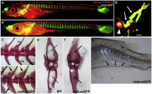
Osteoclast analysis in Rankl transgenic medaka embryos. (a, a’) Live imaging of macrophages (mpeg:mCherry) and osteoclasts (ctsk:GFP) in rankl:HSE:CFP transgenic medaka embryos at 1 (a) and 3 (a’) days after Rankl induction by heat-shock at 9 days post fertilization. Upon Rankl induction, mpeg:mCherry positive macrophages accumulate at the vertebral column and differentiate into ctsk:GFP expressing osteoclasts. Note that GFP positive cells in the head and caudal fin are cathepsin K expressing cells of unknown identity. (b) High magnification view of vertebral neural arch region showing two macrophages that have differentiated into osteoclasts (ctsk:GFP and mpeg:mCherry double-positive; arrows), and a macrophage that has engulfed a dying osteoclast (arrowhead). Asterisk indicates a non-differentiated macrophage. (c, d) Osteopetrosis phenotype in an osteoclast-deficient, ctsk-mEGFP transgenic medaka, expressing a membrane-localized version of GFP in osteoclasts. Lateral views of Alizarin red stained mineralized matrix in the vertebral column of a wild type (c) and ctsk:mEGFP transgenic medaka (d) at 3 months post fertilization. (e, f) Transverse view of individual vertebra of wild type (e) and ctsk:mEGFP transgenic medaka (f). Regions of over-ossification are indicated with arrows. (g) Trunk of 3 week old ctsk:mEGFP fish; dislocated blood vessels are indicated by arrows. Images (A-B) are courtesy of Phan Quang Tien (Phan and Winkler, unpublished), images (C-G) are taken from To et al. (2015). |
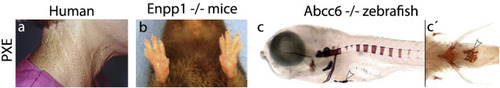
Mouse and zebrafish mutants for Abcc6 and Enpp1 recapitulate some of the over-mineralization seen in Pseudoxanthoma elasticum (PXE) patients. a) PXE patients present areas of mineralization in the skin (taken from the American Osteopathic College of Dermatology). b) Mouse mutants for Enpp1 develop joint stiffness and contractures. In addition, mineralization has been found in the heart and circulatory system, retina and kidneys (Li et al., 2013) (image taken from (Li et al., 2013)). c) Zebrafish mutants for abcc6, resembling human PXE patients in the mineralization in the skin (image taken from (Mackay et al., 2015)). |
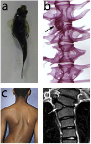
Scoliosis defects found in human patients and in zebrafish. (a) tbx6 mutants (fss) can develop scoliosis. In some of the cases the curvature in the axis is due to a defect in vertebral patterning (b). Patients with scoliosis (c) can also have the same defect in vertebral patterning (d). Image c taken from the website Vikalp physiotherapy clinic. Image d was taken from (Johal et al., 2016). |
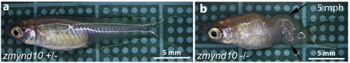
Spine curvature phenotype in the medaka mutant zmynd10 (Kobayashi et al., 2017) at 5 mph. Arrows point at the sides of axis bending. Images were kindly provided by Daisuke Kobayashi. Mph, months post-hatching. |
Reprinted from Developmental Biology, 457(2), Lleras-Forero, L., Winkler, C., Schulte-Merker, S., Zebrafish and medaka as models for biomedical research of bone diseases, 191-205, Copyright (2019) with permission from Elsevier. Full text @ Dev. Biol.