
- Title
-
Bifunctional Small Molecules Enhance Neutrophil Activities Against Aspergillus fumigatus in vivo and in vitro
- Authors
- Jones, C.N., Ellett, F., Robertson, A.L., Forrest, K.M., Judice, K., Balkovec, J.M., Springer, M., Markmann, J.F., Vyas, J.M., Warren, H.S., Irimia, D.
- Source
- Full text @ Front Immunol
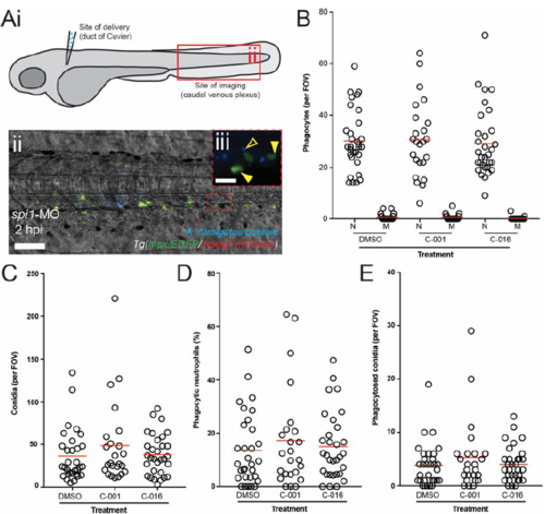
Bifunctional compounds do not enhance phagocytosis of conidia by wild-type zebrafish neutrophils. A) Diagram of a 72 hpf zebrafish larva indicating the site of inoculation and the site of analysis (i). (ii) Collapsed z-stack (maximum intensity) representative image showing conidia (blue, Hoechst) and neutrophils (green, GFP) in the caudal venous plexus of Tg(mpx:GFP/mpeg1:mCherry) embryos injected with spi1-MO at the one-cell stage and infected with A. fumigatus conidia at 72 hpi. Scale: 100 μm. iii) highermagnification of neutrophils and conidia indicating extracellular conidia (open yellow arrowhead) and examples of phagocytosis by neutrophils (filled yellow arrowheads). Scale: 20 μm. B) Graph shows neutrophil (N) and macrophage (M) counts in each caudal venous plexus field of view (FOV) for spi1-MO morphant larvae injected with treated and control conidia. C) Graph shows conidia counts per FOV for each treatment group. D) Graph shows the percent of neutrophils containing conidia at 2 hpi for each treatment group. E) Graph shows the number of phagocytosed conidia per FOV at 2 hpi for each treatment group. Each point represents an infected larva. |
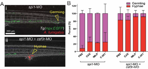
Treatment with bifunctional compounds does not enhance the neutrophil-dependent suppression of fungal hyphae in wild-type zebrafish. A) Representative images of spi1 (i) and spi1/csf3r (ii) morpholino-injected zebrafish larvae infected with A. fumigatus at 1 dpi. Different A. fumigatus growth forms are indicated by open yellow arrowheads. B) Graph showing the proportion of surviving infected embryos with germinating conidia or fully-developed hyphae at 1 dpi. Error bars: Mean + SEM. N = 10 larva per group per experiment, data collated from ≥ 3 experiments |
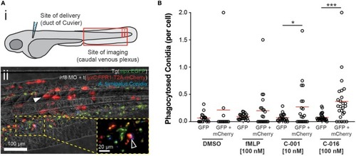
Bifunctional compounds enhance phagocytosis of |