FIGURE SUMMARY
Title

Morphogenesis and axis specification occur in parallel during optic cup and optic fissure formation, differentially modulated by BMP and Wnt

Authors
Eckert, P., Knickmeyer, M.D., Schütz, L., Wittbrodt, J., Heermann, S.
Source
Full text @ Open Biol.

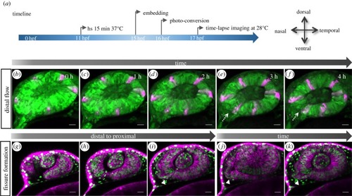
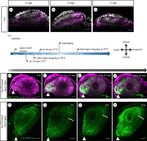
Transformation of the optic vesicle into the optic cup. (a) Timeline of experimental procedure (for bf) and orientation of the eye. (bf) Distal flow and fissure generation. (b) Photo-converted domains (magenta) in the lens-averted layer move over the distal rims into the lens-facing domain (cf). During this process, the fissure is induced (dotted arrow). (gi) The optic fissure is generated from distal to proximal. (ik) The optic stalk (arrowhead) is connected to the nasal lens-averted domain of the optic vesicle, imaging starts (gj) at 19 hpf; scale bar, 25 µm.

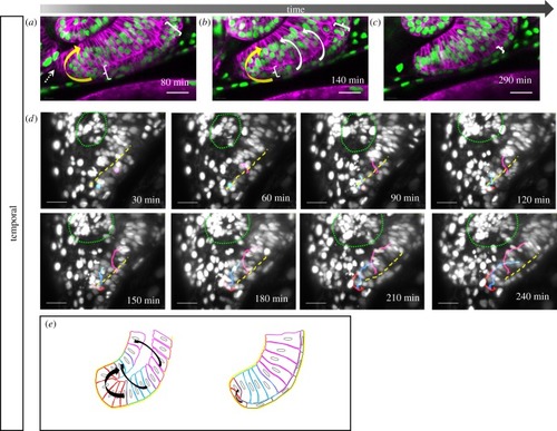
Development of the temporal fissure margin. (ae) Close up of the developing temporal ventral optic cup domain, (ac) labelled with tg(bact:H2BGFP) and tg(bact:lyntdTomato), cells from the lens-averted layer of the optic cup (a, bracket) are flowing over the distal rims (a, b white arrows) and in a perpendicular direction (a, b, yellow arrow) over the ventral margin into the lens-facing layer (6 fish in 4 experiments). (c, bracket) Cells in the lens-averted layer (RPE domain) flatten and obtain RPE cell shape. Lateral view, nasal to the left; scale bar, 25 µm. (d) Mosaic nuclear labelling (H2BGFP mRNA injection), maximum projection of 70 optical sections corresponding to 70 µm (z-spacing was 1 µm). Tracking of single cells (from one eye) moving over the distal (magenta and blue, n = 2 for both, respectively) and the ventral distal (red, n = 4) rim (dashed yellow line), respectively, into the lens-facing layer of the prospective neuroretina. Lens marked with green dotted line. Lateral view, nasal to the left; scale bar, 25 µm. (e) Scheme of temporal fissure margin development. Cells move over the distal (magenta), the distal ventral (blue) rims and via a ventral perpendicular flow (red) over the ventral rim (black arrows) from the lens averted into the lens-facing layer.

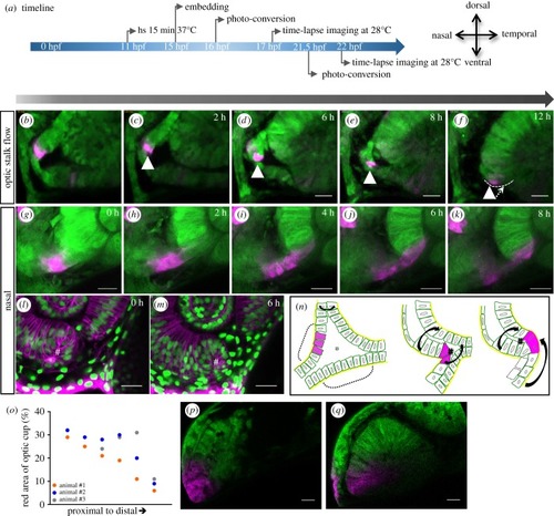
Development of the nasal fissure margin. (a) Timeline of experimental procedure and orientation of the eye (timeline for bf displayed on top of the arrow, for gk below). (bf and gk) Close ups of the developing nasal ventral optic cup labelled with tg(HSP70::kaede), photo-converted cells (magenta) derived from the optic stalk translocate from the optic stalk into the nasal fissure margin (12 fish in 6 experiments; not all of them are kaede experiments). Lateral view, nasal to the left, scale bar: 25 µm. (lm) In distal domains, a turbulence or vortex of cells can be appreciated (hash), in this domain, two flow movements collide, one over the distal rim, one from the optic stalk. Cells remaining in the RPE domain flatten and thus obtain RPE cell shape (labelled by tg(bact:H2BGFP) and tg(bact:lyntdTomato)). (n) Scheme of nasal fissure margin development. In proximal regions of the early optic cup, the optic stalk consists of two layers (dashed brackets), which are connected to the lens-averted domains of the optic cup (see also figure 1g) and border the optic ventricle (asterisk). Over time, the upper layer is moving into the optic fissure margin. The distal and optic stalk flow movements are indicated by black arrows. The twist of the optic stalk could easily be driven by the distal flow on the nasal side which is flowing over the optic stalk. Eventually, stalk-derived cells are integrated via the nasal fissure margin. (o) Optic stalk contribution to the ventral neuroretina (stalks of three animals were converted and the red area was measured separately). (p) Optic vesicle from animal #2 before fissure development. Parts of the optic stalk were converted. (q) Optic cup from animal #2 after fissure development. Photo-converted cells moved into the ventral area of the optic cup. Scale bar, 25 μm.

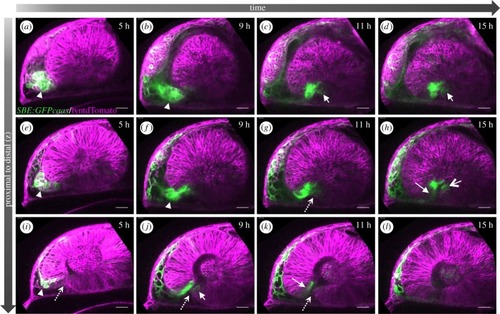
TGFβ-signalling-positive cells are secondarily added to the optic fissure margins. (al) Four-dimensional dataset of the developing optic cup, TGFβ reporter (green), cell membranes (lyntdTomato, magenta). Presented are three different optical planes (top to bottom) over time (left to right). TGFβ reporter activity in the optic stalk (a,e,i, arrowhead). TGFβ-signalling-positive cells move from the optic stalk into the optic cup (ck, arrow) (8 fish in 4 experiments; 3 fish in 2 experiments). The optic stalk is predominantly connected to the nasal optic cup (a,b,e,f). At the end of the flow, TGFβ-signalling-positive cells populate the most proximal domain of the optic fissure (c,d,f,g) (3 fish in 2 experiments). From here, these cells also populate the temporal fissure margin (c,d,f,g,h,j). The optic fissure is marked with a dotted arrow. Lateral view, nasal to the left; scale bar, 25 µm.

Induced expression of bmp4 hampers optic fissure formation. In situ hybridizations for fsta (ac) for 13, 15 and 17 hpf in WT embryos. fsta is expressed, from temporal to the ventral transition zone to the optic stalk. (d) Timeline of the experimental procedure and orientation of the eye, heat shocks (hs) performed at 17 hpf are displayed on top of the arrow, hs performed at 13 hpf below the arrow. (eh) Lateral view of optic cup development in the tg(hsp70:bmp4) background, visualized by lyntdTomato (mRNA), tg(SBE:GFPcaax). bmp4 induced at 17 hpf hampers proximal optic fissure morphogenesis (10 fish in 1 experiment). The optic stalk is in continuation to the lens-averted domains of the developing optic cup (e, arrowhead). Asterisk marks the optic ventricle. In temporal and dorsal regions, the lens-averted layer is being integrated into the optic cup (f–h). Cells from the lens-averted layer of the optic cup are not properly integrated into the lens-facing domain. The connection of the optic stalk to the lens-averted domain is maintained and the optic fissure is not formed in the proximal domain. The TGFβ signalling activity can be seen in the optic cup, in the absence of orderly tissue dynamics. See figure 4 as control. (il) Lateral view of optic cup development in the tg(hsp70:bmp4) background, visualized by tg(rx2:GFPcaax). bmp4 induced at 13 hpf results in an absence of the optic fissure (4 fish in 1 experiment). The optic stalk is misshaped (arrowhead). On the temporal side, a persisting lens-averted domain is visible (brackets jl). Scale bar, 25 μm. See electronic supplementary material, figure 5 supplement 1 O–X as control.

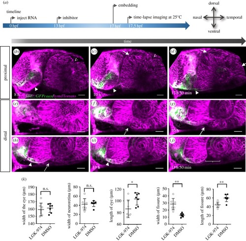
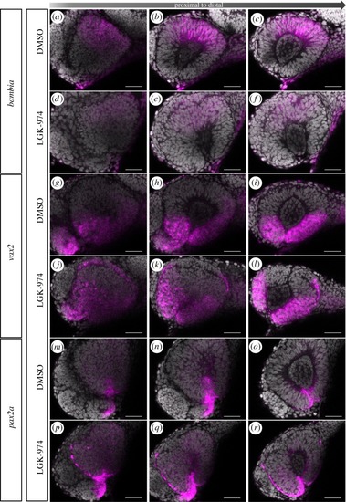
Wnt-signalling inhibition affects optic cup morphogenesis and prevents TGFβ-signalling positive cells from entering the ventral part of the optic cup. (a) Timeline of the experimental procedure, and orientation of the eye. (bj) Four-dimensional dataset of a developing optic cup. TGFβ reporter (green); cell membranes (lyntdTomato, magenta). One optical section in a proximal (bd) and one in a distal region (ej) over time (left to right). TGFβ-signalling-positive cells are located in the misshaped optic stalk/forebrain (arrowhead). Few TGFβ-signalling-positive cells reach the nasal ventral part of the developing optic cup (h,i, arrowhead). The dorsal fissure (bc, marked with v) seems to close over time. Ectopic domains of the presumptive neuroretina can be seen in the lens-averted dorsal domain (cd, arrows). Even though TGFβ-positive cells do not move into the eye, a nasal fissure margin is visible. On the temporal side, the ventral perpendicular flow seems corrupted, affecting the formation of the temporal fissure margin. The distal flow in the ventral domains, both nasal and temporal, seems unaffected resulting in an optic fissure being visible in distal domains (i,j brackets). A dotted arrow indicates where the fissure will open (5 fish in 1 experiment, 3 TGFB reporter, 2 WT). Scale bar, 25 µm. (k) Quantification of distinct morphological parameters of LGK-974-treated embryos versus DMSO-treated embryos. *p < 0.05; **p < 0.005.

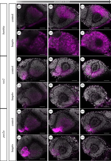
Induced bmp4 expression affects moprphogenesis and axis specification. In situ hybridizations for bambia (af), vax2 (gl) and pax2a (mr) in tg(hsp70:bmp4) (df, jl, pr) and control embryos (ac, gi, mo) at 24 hpf after heat shock at 17 hpf. Note the extended bambia expression domain within the entire optic cup resulting from bmp4 induction, compared to the dorsal expression domain in the control. Vax2 expression is reduced after bmp4 induction compared to the control. Bmp4 induction also results in a reduced pax2a expression domain within the optic cup, while the pax2a expression domain in the optic stalk is enlarged. Lateral view, nasal to the left; scale bar, 25 µm. For each condition, eight embryos were used; four to five of them were imaged.

Inhibition of Wnt-signalling affects morphogenesis. In situ hybridizations for bambia (af), vax2 (gl) and pax2a (mr) on porcupine inhibitor (LGK-974)-treated embryos (df, jl, pr) and DMSO control embryos (ac, gi, mo) at 24 hpf. Inhibitor treatment started at 13 hpf. Wnt-signalling inhibition is not affecting the expression domain of bambia but reducing expression intensity. Wnt-signalling inhibition results in ectopic expression of vax2, which can be found in the lens-averted layer of the optic cup. Wnt-signalling inhibition also results in a reduced pax2a expression domain within the optic cup, while the pax2a expression domain in the optic stalk is enlarged and extending into the nasal lens-averted domain. Lateral view, nasal to the left; scale bar, 25 µm. For each condition, eight embryos were used; four to five of them were imaged.

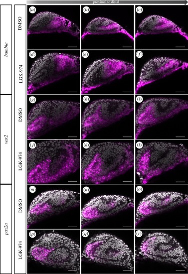
Inhibition of Wnt-signalling does not affect dorsal ventral axis specification. In situ hybridizations for bambia (af), vax2 (gl) and pax2a (mr) on LGK-974-treated embryos (df, jl, pr) and DMSO control embryos (ac, gi, mo) at 17 hpf. Inhibitor treatment started at 13 hpf. No change of the expression pattern was found for bambia and vax2. Pax2a, however, was found expressed ectopic within the nasal lens-averted layer resulting from Wnt-signalling inhibition. Lateral view, nasal to the left; scale bar, 25 µm. For each condition, eight embryos were used; four to five of them were imaged.

Acknowledgments
This image is the copyrighted work of the attributed author or publisher, and ZFIN has permission only to display this image to its users. Additional permissions should be obtained from the applicable author or publisher of the image. Full text @ Open Biol.