
- Title
-
Countershading in zebrafish results from an Asip1 controlled dorsoventral gradient of pigment cell differentiation
- Authors
- Cal, L., Suarez-Bregua, P., Comesaña, P., Owen, J., Braasch, I., Kelsh, R., Cerdá-Reverter, J.M., Rotllant, J.
- Source
- Full text @ Sci. Rep.
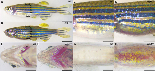
Adult dorso-ventral countershading pattern is disrupted in asip1K.O.. Lateral (A,B), anterior-lateral (C,D), ventral head (E,F) and ventral belly (G,H) views of 180 dpf WT and asip1K.O. zebrafish. (A,B) The pigment pattern of WT zebrafish is a striped pigment pattern with dark stripes and light interstripes. Each dark stripe is named with a code: two primary stripes are called 1D and 1 V, and the two secondary stripes are named 2D and 2 V. The asip1K.O. display an extra 3 V dark stripe. The asip1K.O. phenotype is characterized by a darker belly than WT. (C,D) The striped pigment pattern was almost unaltered in asip1K.O. fish, except that the 2 V stripe is wider than in WT, and the ventral dark stripe 3 V is better developed anteriorly. The darker belly of asip1K.O. compared to WT sibling fish is clearly evident. (E,F) In WT, melanophores are infrequent around the jaws and branchiostegals; however, branchiostegal, jaw and operculum regions are clearly hyperpigmented in asip1K.O.. (G,H) Melanophores are virtually absent in WT belly; thus, WT ventral region shows bright white color as a result of high number of iridophores in the abdominal wall. However, asip1K.O. shows a consistent hyperpigmentation, with many melanophores and xanthophores in the ventral skin; the abdominal wall also seems to be affected, with reduced extent of iridophores and looking much yellower than WT. Scale bar: (A,B) 5 mm, (C–H) 2 mm. Abbreviation: br, branchiostegal.
PHENOTYPE:
|
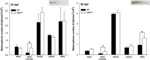
Dorsal-ventral distribution of melanophores during metamorphosis. (A) Distribution and number of melanophores in 15 dpf WT and asip1K.O. fish. At this stage, asip1K.O. already shows significantly higher number of melanophores in the ventral view of the head. (B) Distribution and number of melanophores in WT and asip1K.O. 30 dpf fish. At this stage, asip1K.O. shows significantly higher number of melanophores in the ventral view of the head, but also in the belly. Data are the mean ± SEM, n = 7. Asterisks indicate significant differences between WT and asip1K.O. fish. Scale bar: (A) 200 μm, (B) 500 μm.
PHENOTYPE:
|

Quantitation of dorsal-ventral distribution of melanophores and xanthophores in adult WT and asip1K.O.fish. (A) Lateral view of zebrafish showing the body regions selected for melanophore and xanthophore count. (B) Ventral view of the WT and asip1K.O. 210 dpf zebrafish fish belly. (C) Distribution and number of melanophores in WT and asip1K.O. 60 dpf fish. At this stage, asip1K.O. shows a significantly higher number of melanophores in the black stripe 2 V, ventral head and belly. (D) Number of xanthophores in the dorsal and ventral skin of WT and asip1K.O. 60 dpf fish. At this stage, asip1K.O. shows a significantly higher number of xanthophores in the belly region. (E) Distribution and number of melanophores in WT and asip1K.O. 210 dpf fish. At this stage, asip1K.O. shows significantly higher number of melanophores also in black stripe 2 V, 3 V, ventral head and belly. (F) Number of xanthophores in dorsal and ventral skin of WT and asip1K.O. 210 dpf fish. These fish showed highly significant higher number of xanthophores in belly region than WT. Data are the mean ± SEM, n = 7. Asterisks indicate significant differences between WT and asip1K.O. fish. Scale bar (A,C,E) 1 mm, (B) 100 μm.
PHENOTYPE:
|
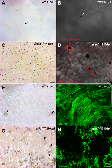
Detailed visualization of ventral pigment cells in WT and asip1 mutants. (A) Ventral view of 210 dpf WT belly. (B) Belly of 210 dpf WT fish carrying Tg(Kita:GalTA4;UAS:mCherry) (labels melanophores) transgene shows no melanophores in ventral skin. (C) Ventral view of 210 dpf asip1K.O. belly. (D) Belly of 210 dpf asip1K.O. fish carrying Tg(Kita:GalTA4;UAS:mCherry) transgene shows high number of melanophores in ventral skin. (E) Internal view of 210 dpf WT abdominal wall shows a white sheet of iridophores with few internal melanophores (black arrow). (F) Abdominal wall of 210 dpf WT fish carrying Tg(TDL358:GFP) (labels iridophores and glia) transgene displays a uniform and continuous sheet of iridophores. (G) Internal view of 210 dpf asip1K.O. abdominal wall shows a disrupted and discontinuous sheet of iridophores with high number of melanophores (black arrow) and some xanthophores (orange arrow). (H) Abdominal wall of 210 dpf asip1K.O. fish carrying Tg(TDL358:GFP)transgene exhibits a broken sheet of iridophores. Scale bars: 100 μm. |
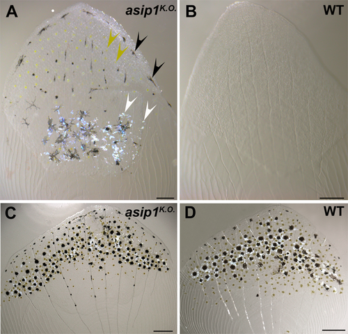
Adult asip1K.O. ventral scales displayed a dorsalized color pattern. (A) 210 dpf asip1K.O. ventral scales exhibit a pattern of melanophores (black arrowheads), xanthophores (yellow arrowheads) and also iridophores (white arrowheads). (B) 210 dpf WT ventral scale does not exhibit any chromatophores. (C,D) 210 dpf WT and asip1K.O. dorsal scales exhibit a similar pattern of melanophores, xanthophores and iridophores. Scale bars: 100 μm. PHENOTYPE:
|
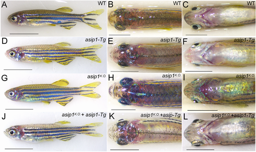
Functional rescue of CRISPR-mediated asip1 mutation. Lateral (A,D,G,J), dorsal (B,E,H,K) and ventral-belly (C,F,I,L) views of 160 dpf WT, asip1-Tg, asip1K.O., and asip1K.O; asip1-Tg zebrafish. The pigment pattern of WT zebrafish shows (A) normal striped pattern, (B) dark dorsum and (C) light belly. The pigment pattern of asip1-Tg fish shows (D) almost normal striped pattern, although dark stripe 2D??? is rather thinner???, (E) hypopigmented dorsum and (F) light belly. The pigment pattern of asip1K.O. fish shows (F) almost normal striped pattern, but with dark stripes 2 V and 3 V more developed than WT fish, (H) pigmented dorsum similar to WT and (I) hyperpigmented belly compared to WT. The asip1K.O + asip1 − Tg phenotype shows a phenotype similar to the asip1 − Tg zebrafish, except that dark stripe 2D is more prominent. Scale bar: 5 mm.
PHENOTYPE:
|