
- Title
-
Dual Inhibition of GSK3β and CDK5 Protects the Cytoskeleton of Neurons from Neuroinflammatory-Mediated Degeneration In Vitro and In Vivo
- Authors
- Reinhardt, L., Kordes, S., Reinhardt, P., Glatza, M., Baumann, M., Drexler, H.C.A., Menninger, S., Zischinsky, G., Eickhoff, J., Fröb, C., Bhattarai, P., Arulmozhivarman, G., Marrone, L., Janosch, A., Adachi, K., Stehling, M., Anderson, E.N., Abo-Rady, M., Bickle, M., Pandey, U.B., Reimer, M.M., Kizil, C., Schöler, H.R., Nussbaumer, P., Klebl, B., Sterneckert, J.L.
- Source
- Full text @ Stem Cell Reports
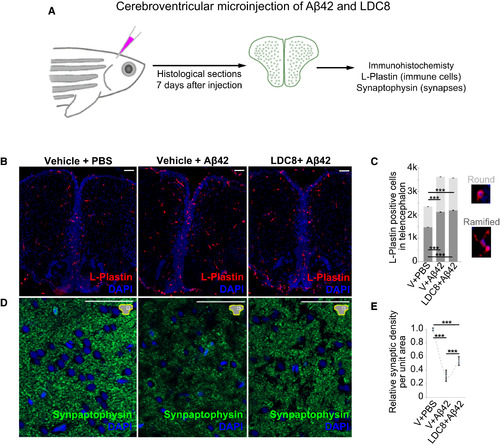
LDC8 Reduces the Synaptic Degeneration in an Adult AD Model in Zebrafish (A) Graphic representation of the cerebroventricular microinjection and experimental scheme in adult zebrafish brain. (B) Immunohistochemistry for L-plastin (immune cells) in fish brains injected with vehicle and PBS, vehicle and Aβ42, and LDC8 and Aβ42. (C) Quantification graph for the numbers of active (round) and resting (ramified) immune cells after three injection conditions. Insets next to the graph indicate typical morphology. (D) Immunohistochemistry for synaptophysin (synaptic connections) in fish brains injected with vehicle and PBS, vehicle and Aβ42, and LDC8 and Aβ42. (E) Quantification graph for the relative synaptic density. Aβ42 significantly reduces the synaptic connections while LDC8 provides a significant increase in the surviving synapses. ∗∗∗p < 0.001. Scale bars, 50 μm. See also Figure S7. |

ZFIN is incorporating published figure images and captions as part of an ongoing project. Figures from some publications have not yet been curated, or are not available for display because of copyright restrictions. |