
- Title
-
Using zebrafish larval models to study brain injury, locomotor and neuroinflammatory outcomes following intracerebral haemorrhage
- Authors
- Crilly, S., Njegic, A., Laurie, S.E., Fotiou, E., Hudson, G., Barrington, J., Webb, K., Young, H.L., Badrock, A.P., Hurlstone, A., Rivers-Auty, J., Parry-Jones, A.R., Allan, S.M., Kasher, P.R.
- Source
- Full text @ F1000Res
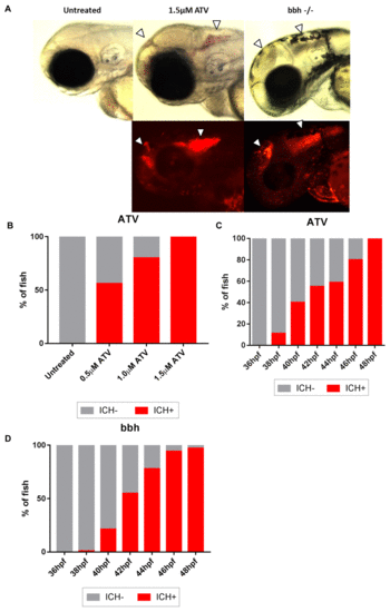
Atorvastatin (ATV)-induced and bubblehead (bbh) mutant intracerebral haemorrhage (ICH) show comparable models of brain-specific bleeding. (A) Brain-specific bleeds were observed in both ATV and bbh models maintained on the transgenic gata1:DsRed reporter background using both brightfield (top panels) and fluorescence (bottom panels) microscopy. Bleeds formed in both forebrain and mid-hindbrain regions, as described by others (Eisa-Beygi et al., 2013) (arrows denotes haemorrhages). Bleeds in bbh mutants are frequently associated with severe cranial oedema making blood pooling more disperse. Original magnification, x20. (B) ATV treatment causes ICH to occur in a dose-dependent manner. (C) Timeline of ICH development in ATV-treated and untreated embryos and (D) bbh homozygotes. |
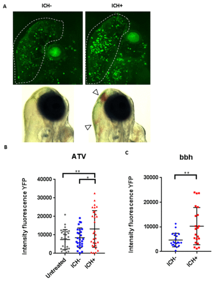
Intracerebral haemorrhage (ICH) in zebrafish larvae results in a quantifiable brain injury. (A) Representative images of the brain injury phenotype in ICH+ larvae (right panels), in comparison to ICH- siblings (left panels), at 72 hpf. Bright-field images (bottom panels) demonstrate the presence of brain bleeds (arrows) in ICH+ larvae. Fluorescent microscopy was performed to visualise cell death in the ubiq:secAnnexinV-mVenus reporter line (top panels). Clusters of dying cells were observed in peri-haematomal regions. Images were cropped to brain only regions and analysed for total green fluorescence intensity in round particles bigger than 30 pixels in diameter (white line). (B) Quantification of fluorescent signal in the brains of untreated, ICH- and ICH+ larvae obtained through the ATV model (n=12 per group; 3 independent replicates) at 72 hpf. Significant differences were observed when comparing ICH+ with untreated (**p=0.004) and with ICH- (*p=0.03) siblings. (C) Quantification of fluorescent signal as a read out for annexinV binding in the brains of ICH- and ICH+ larvae obtained through the bubblehead (bbh) model (n=12 per group; 2 independent replicates) at 72 hpf. A significant difference in mVenus fluorescence was observed between ICH+ and ICH- age-matched siblings (**p=0.002). Original magnification, x20. EXPRESSION / LABELING:
PHENOTYPE:
|

ZFIN is incorporating published figure images and captions as part of an ongoing project. Figures from some publications have not yet been curated, or are not available for display because of copyright restrictions. PHENOTYPE:
|
|
ZFIN is incorporating published figure images and captions as part of an ongoing project. Figures from some publications have not yet been curated, or are not available for display because of copyright restrictions. PHENOTYPE:
|
|
ZFIN is incorporating published figure images and captions as part of an ongoing project. Figures from some publications have not yet been curated, or are not available for display because of copyright restrictions. |